. 2010 Jun;24(6):1838-51.
doi: 10.1096/fj.09-151639. Epub 2010 Jan 22.

Vimentin induces changes in cell shape, motility, and adhesion during the epithelial to mesenchymal transition

Affiliations

Vimentin induces changes in cell shape, motility, and adhesion during the epithelial to mesenchymal transition

Melissa G Mendez et al. FASEB J. 2010 Jun.

Abstract

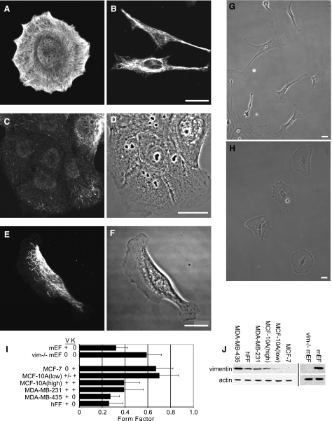
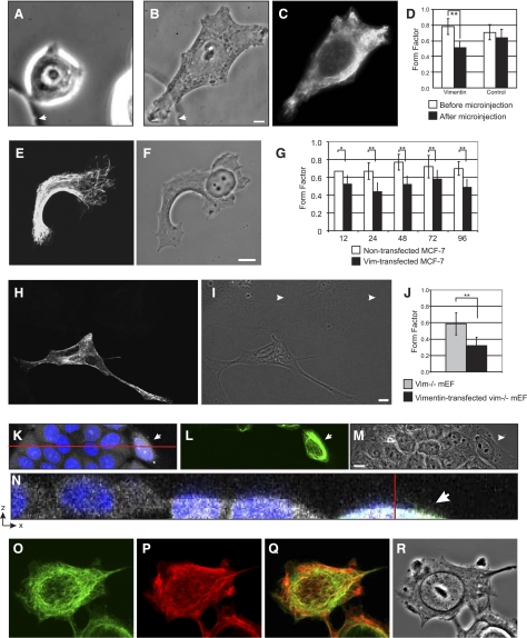
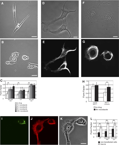
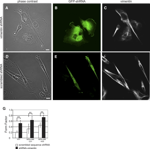
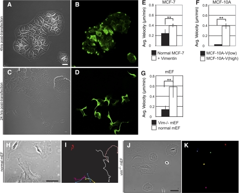
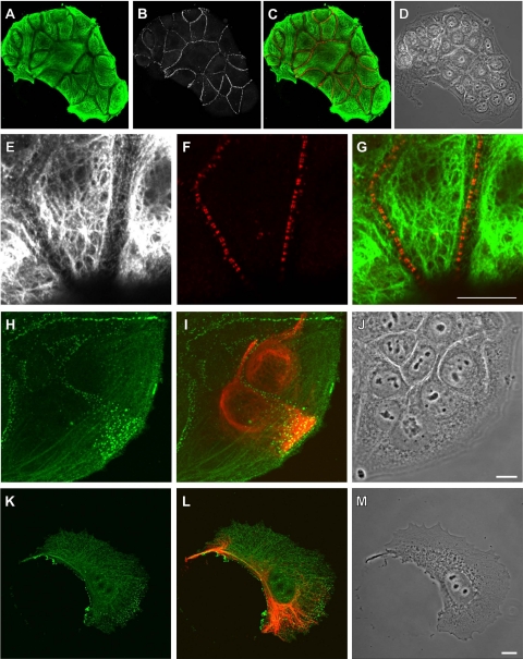
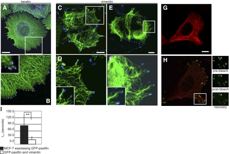
Vimentin is used widely as a marker of the epithelial to mesenchymal transitions (EMTs) that take place during embryogenesis and metastasis, yet the functional implications of the expression of this type III intermediate filament (IF) protein are poorly understood. Using form factor analysis and quantitative Western blotting of normal, metastatic, and vimentin-null cell lines, we show that the level of expression of vimentin IFs (VIFs) correlates with mesenchymal cell shape and motile behavior. The reorganization of VIFs caused by expressing a dominant-negative mutant or by silencing vimentin with shRNA (neither of which alter microtubule or microfilament assembly) causes mesenchymal cells to adopt epithelial shapes. Following the microinjection of vimentin or transfection with vimentin cDNA, epithelial cells rapidly adopt mesenchymal shapes coincident with VIF assembly. These shape transitions are accompanied by a loss of desmosomal contacts, an increase in cell motility, and a significant increase in focal adhesion dynamics. Our results demonstrate that VIFs play a predominant role in the changes in shape, adhesion, and motility that occur during the EMT.


Figures

Figure 1.
Figure 1.
VIF expression levels correlate with cell shape. A, B) MCF-7 epithelial cells express only KIFs (A), and MDA-MB-435 mesenchymal cells express only VIFs (B). C–F) MCF-10A-V (low) cells contain mainly vimentin particles and squiggles (C, D), while MCF-10A-V (high) cells contain more extensive VIF networks (E, F). G) Normal mEFs. H) Vim−/−mEFs. I) FF of each cell line. J) Immunoblots of whole-cell extracts of the different cell lines. Scale bars = 10 μm.
Figure 2.
Figure 2.
Vimentin induces epithelial cells to adopt mesenchymal shapes. A–D) An MCF-7 cell immediately before microinjection (A) and 5 h postinjection after processing for immunofluorescence (B, C; vimentin). Arrow indicates fiducial mark on gridded coverslip. At 2–5 h after injection, FFs reflect more mesenchymal shapes(D). E–G) Following transient transfection, MCF-7 cells that contain VIF networks (E) adopt mesenchymal shapes (F). FF of these cells is significantly less than that of control cells (compare to Fig. 1A) on the same coverslip (G). H–J) Vim−/−mEFs expressing VIFs (H) appear like normal mEFs (I, J); arrowheads indicate nontransfected vim−/−mEFs. K–N) MCF-7 cells expressing VIFs are shorter than nontransfected controls. Arrowheads indicate the same cell expressing vimentin. Blue, Hoechst (K, N); green, VIFs (L, N); white, Vybrant dye (M, N). O, P) Expression of KIFs (O) is retained by MCF-7 cells expressing VIFs (P). Q) Overlay of O, P. R) Phase contrast. Bars = 10 μm.
Figure 3.
Figure 3.
Reorganization of VIFs induces mesenchymal cells to adopt epithelial-like cell shapes. AC) MDA-MB-435 cells (A) were exposed to nocodazole for 1–24 h (B; 24 h). FF analysis (C) shows that nocodazole-treated cells become more circular in shape. D–H). VIFs in transfected MCF-7 cells (D, phase contrast; E, immunofluorescence with vimentin antibody of the same cells) reorganize into a perinuclear cap following exposure to nocodazole (F, phase contrast; G, immunofluorescence of the same cell with vimentin antibody). Concurrent with this VIF reorganization, MCF-7 cells containing VIFs revert to more circular shapes (F–H). In contrast, the shapes of normal MCF-7 do not respond to nocodazole treatment (H). I–K) Following transfection with the Vim1A-GFP dominant-negative mutant, VIFs in MDA-MB-435 cells reorganize into perinuclear aggregates. Same field of fixed cells shows Vim1A-GFP(I), immunofluorescence with vimentin antibody (J), and phase contrast (K). L) FF analysis of Vim1A-GFP expressing cells. Scale bars = 10 μm.
Figure 4.
Figure 4.
Vimentin silencing induces mesenchymal cells to adopt epithelial-like cell shapes. A–C) MDA-MB-435 cells silenced with GFP-tagged vimentin shRNA adopt more circular shapes: A) phase contrast; B) immunofluorescence with GFP antibody; C), vimentin. Vimentin that remains in these cells assembles into disorganized short filaments (C). D–F) Cells transfected with GFP-tagged scrambled-sequence control shRNA do not change shape: D) phase contrast; E) immunofluorescence with GFP antibody; F), vimentin. G) Mesenchymal cells respond similarly to vimentin silencing regardless of whether KIFs are also present. Scale bars = 10 μm.
Figure 5.
Figure 5.
Motility is enhanced in VIF-expressing epithelial cells. A, B) Four hours following transfection with vimentin cDNA, vimentin-expressing cells are distributed throughout MCF-7 colonies: A) phase contrast; B) vimentin. In the first 2–4 h after transfection (A), vimentin is expressed in most cells. At this time, some cells show extensive expression throughout the cytoplasm; others contain small regions, sometimes concentrated in peripheral regions (B). By ∼6 h post-transfection, obvious vimentin expression is detected in ∼12% of the cells. Although we cannot explain the transient appearance of vimentin in so many cells, it appears that these changes are inherent to the process of transfection. C, D) By 24 h post-transfection, most cells containing VIFs are located at the peripheries of colonies or have moved away from colonies: C) phase contrast; D) vimentin; fixed and processed for immunofluorescence. E–G) Live-cell imaging reveals that MCF-7 cells expressing VIFs move faster than nontransfected cells (E); and that MCF-10A-V (high) cells move faster than MCF-10A-V (low) cells (F). In addition, wild-type mEFs move much faster than vim−/− mEFs (G). H–K) Relative motility is evidenced by the tracings of the positions of nuclei over 6 h (H, I) and 4 h, respectively. It is evident that Vim−/− mEFs are virtually nonmotile (J, K). See Supplemental Movies 1 (normal mEFs) and 2 (vim−/− mEFs). Scale bars = 10 μm (A, C); 100 μm (H, J).
Figure 6.
Figure 6.
Desmosomes are internalized in MCF-7 cells expressing VIFs. A–D) Under normal conditions, MCF-7 cells within a colony are linked by desmosomes, as seen by immunofluorescence: A) keratin; B) desmoplakin; C) overlay (desmoplakin, red); D), phase contrast. E–G) Higher magnification views: E) keratin; F) desmoplakin; G) overlay. H–J) Desmosomes (H, green) appear to be undergoing internalization in MCF-7 cells containing VIFs (I, red), as visualized by immunofluorescence (J, phase contrast). K–M) Desmosomes (K; desmoplakin, green) are no longer aligned along the edges of MCF-7 cells that contain VIFs (L, red; M, phase contrast) and are no longer in contact with other cells. Scale bars = 10 μm.
Figure 7.
Figure 7.
Properties of paxillin are altered in epithelial cells expressing VIFs. A) Immunofluorescence image showing that KIFs in MCF-7 cells extend toward FAs (paxillin, blue). B) Area boxed in A and inset at higher magnification. C–F) Following transfection with vimentin cDNA and processing for immunofluorescence, VIFs are seen to extend toward and become closely associated with FAs (paxillin, blue). This occurs soon after transfection (C, D) and persists as VIF assembly progresses (E, F). Panels D, F and insets show areas boxed in C, E at higher magnification. G) To examine FA dynamics, cells cotransfected with cDNA for wild-type vimentin, RFP-vimentin, and GFP-paxillin were observed live. H) Image of more basal focal plane as the same cell in G. Region indicated by the box in H is seen at higher magnification in the three images to the right, before and after FRAP analysis of a photobleached focal contact. I) The t1/2 to recovery from photobleaching decreases 4-fold in MCF-7 cells expressing VIFs. Scale bars = 10 μm.

Similar articles

Cited by

References

    1. Hay E D. The mesenchymal cell, its role in the embryo, and the remarkable signaling mechanisms that create it. Dev Dyn. 2005;233:706–720. - PubMed
    1. Steinert P M, Idler W W, Cabral F, Gottesman M M, Goldman R D. In vitro assembly of homopolymer and copolymer filaments from intermediate filament subunits of muscle and fibroblastic cells. Proc Natl Acad Sci U S A. 1981;78:3692–3696. - PMC - PubMed
    1. Franke W W, Schiller D L, Hatzfeld M, Winter S. Protein complexes of intermediate-sized filaments: melting of cytokeratin complexes in urea reveals different polypeptide separation characteristics. Proc Natl Acad Sci U S A. 1983;80:7113–7117. - PMC - PubMed
    1. Acloque H, Adams M S, Fishwick K, Bronner-Fraser M, Nieto M A. Epithelial-mesenchymal transitions: the importance of changing cell state in development and disease. J Clin Invest. 2009;119:1438–1449. - PMC - PubMed
    1. Kim K K, Kugler M C, Wolters P J, Robillard L, Galvez M G, Brumwell A N, Sheppard D, Chapman H A. Alveolar epithelial cell mesenchymal transition develops in vivo during pulmonary fibrosis and is regulated by the extracellular matrix. Proc Natl Acad Sci U S A. 2006;103:13180–13185. - PMC - PubMed

Publication types

LinkOut - more resources