. 2010 Mar 25;115(12):2449-57.
doi: 10.1182/blood-2009-07-234757. Epub 2010 Jan 22.

Heparanase-enhanced shedding of syndecan-1 by myeloma cells promotes endothelial invasion and angiogenesis

Affiliations

Heparanase-enhanced shedding of syndecan-1 by myeloma cells promotes endothelial invasion and angiogenesis

Anurag Purushothaman et al. Blood. .

Abstract

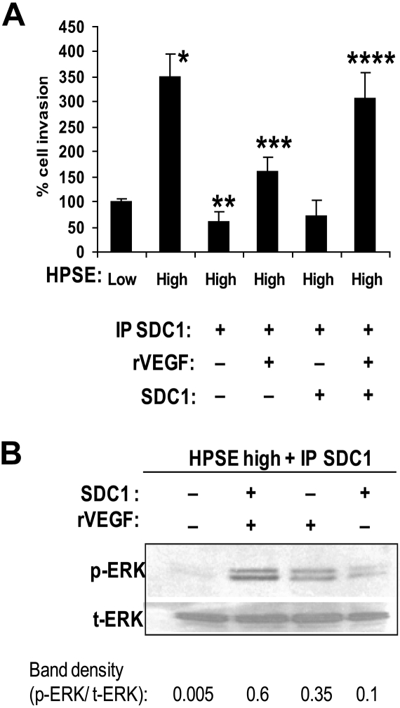
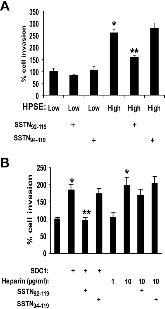
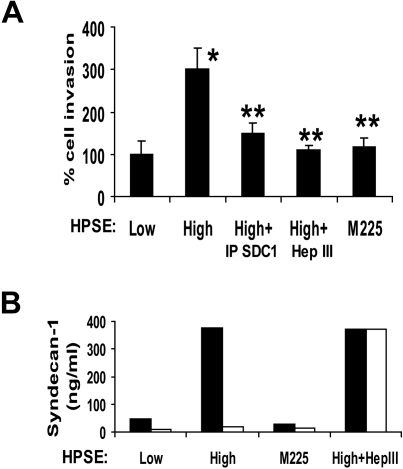
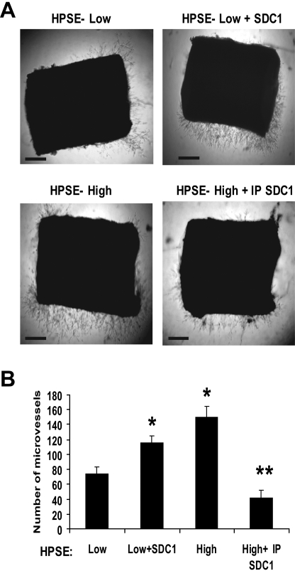
Heparanase enhances shedding of syndecan-1 (CD138), and high levels of heparanase and shed syndecan-1 in the tumor microenvironment are associated with elevated angiogenesis and poor prognosis in myeloma and other cancers. To explore how the heparanase/syndecan-1 axis regulates angiogenesis, we used myeloma cells expressing either high or low levels of heparanase and examined their impact on endothelial cell invasion and angiogenesis. Medium conditioned by heparanase-high cells significantly stimulated endothelial invasion in vitro compared with medium from heparanase-low cells. The stimulatory activity was traced to elevated levels of vascular endothelial growth factor (VEGF) and syndecan-1 in the medium. We discovered that the heparan sulfate chains of syndecan-1 captured VEGF and also attached the syndecan-1/VEGF complex to the extracellular matrix where it then stimulated endothelial invasion. In addition to its heparan sulfate chains, the core protein of syndecan-1 was also required because endothelial invasion was blocked by addition of synstatin, a peptide mimic of the integrin activating region present on the syndecan-1 core protein. These results reveal a novel mechanistic pathway driven by heparanase expression in myeloma cells whereby elevated levels of VEGF and shed syndecan-1 form matrix-anchored complexes that together activate integrin and VEGF receptors on adjacent endothelial cells thereby stimulating tumor angiogenesis.


Figures

Figure 1
Figure 1
Heparanase promotes endothelial cell invasion and ERK phosphorylation. (A) Cells from myeloma cell lines RPMI 8226 or MM.1S were incubated for 24 hours with rHPSE and the conditioned medium collected. Endothelial cells were then placed in the upper chamber of Matrigel invasion chambers and conditioned medium from myeloma cells placed in the lower chamber. After overnight incubation, endothelial cells that invaded through the Matrigel were fixed, stained, and counted. The control was medium that was not conditioned by cells but with addition of rHPSE. Data are mean ± SD of 3 independent experiments. *P < .01 vs medium from cells not exposed to rHPSE. (B) Medium from myeloma cells was collected 24 hours after addition of rHPSE, added to endothelial cells in culture, and cells extracted and prepared for Western blotting for p-ERK and t-ERK. Bands were scanned and densities shown as ratios of p-ERK to t-ERK.
Figure 2
Figure 2
Medium conditioned by heparanase high myeloma cells enhances endothelial invasion via activity of VEGF. (A) Endothelial cells were seeded on the top well of an invasion chamber coated with Matrigel and conditioned medium from either HPSE-low or HPSE-high cells was added to the lower well of the chamber in the presence or absence of a VEGF function blocking antibody. Data are mean ± SD from 3 independent experiments. *P < .001 vs HPSE-low without addition of anti-VEGF. **P < .01 vs HPSE-high. (B) VEGF stimulates ERK activation in endothelial cells. Serum-starved endothelial cells were incubated with conditioned medium from HPSE-high myeloma cells in the presence of VEGF function blocking antibody (Ab-3) or an isotype-matched control antibody. After 15 minutes, whole-cell lysates were prepared and subjected to immunoblotting for p-ERK and t-ERK. (C) VEGF levels are elevated in conditioned medium from HPSE-high cells and a portion of the VEGF associates with syndecan-1. HPSE-low or HPSE-high CAG cells were plated at equal density in complete RPMI medium. After 48 hours, conditioned media were harvested, and the level of VEGF was determined before and after immunodepletion of syndecan-1 (IP SDC1) from the medium (values represent means of triplicate determination ± SD). *P < .001 vs HPSE-low. **P < .001 vs HPSE-high.
Figure 3
Figure 3
Shed syndecan-1 in conditioned medium of HPSE-high myeloma cells facilitates endothelial invasion and ERK phosphorylation. (A) Endothelial invasion is enhanced by shed syndecan-1. An equal number of endothelial cells were seeded on invasion chambers coated with Matrigel and cells allowed to migrate in the presence of medium conditioned by HPSE-low or HPSE-high CAG cells, or medium from HPSE-high cells pretreated with Hep III or IP SDC1 or with medium from cells expressing high levels of an enzymatically inactive form of heparanase (M225). After overnight incubation, cells that invaded through the Matrigel were fixed, stained, and counted. Data are mean ± SD of 3 independent experiments. *P < .01 vs HPSE-low. **P < .05 vs HPSE-high. ***P < .01 vs HPSE-high. (B left panel) Pretreatment of medium with Hep III diminishes ERK activation. Serum-deprived endothelial cells were treated with conditioned medium from HPSE-low or HPSE-high myeloma cells with or without pretreatment with Hep III. At the designated time points, cells were washed and total cell lysates were subjected to immunoblotting with antibodies against p-ERK or t-ERK. (Right panel) ERK signaling is lost after depletion of syndecan-1 from conditioned medium. Medium conditioned by HPSE-high cells was subjected to immunodepletion with control antibody or antibody to syndecan-1. Serum-deprived endothelial cells were treated with this medium for 15 minutes, lysed, and subjected to immunoblotting with antibodies against p-ERK or t-ERK. (C) ERK activation stimulates invasion of endothelial cells. Equal number of endothelial cells along with dimethyl sulfoxide as control, ERK inhibitor PD98059 (50μM), or ERK inhibitor II negative control (50μM) were loaded into the upper compartment of the Matrigel invasion chamber and medium from HPSE-low or HPSE-high cells added to the lower compartment. After overnight incubation, cells that invaded through the Matrigel were fixed, stained, and counted. Data represent mean ± SD of 3 independent experiments. *P < .01 vs HPSE-low without addition of PD98059. **P < .01 vs HPSE-high without addition of PD98059.
Figure 4
Figure 4
Very low levels of heparan sulfate fragments are present in medium from HPSE-high cells. (A) Anion-exchange high-performance liquid chromatography of heparan sulfate fragments eluting at 25 minutes (monosulfated oligosaccharides) and 33 minutes (disulfated oligosaccharides) were detected in the medium conditioned by HPSE-high cells. (B) Heparan sulfate fragments were not detected in medium conditioned by HPSE-low cells. Elution positions of the 2AB-derivatives of standard unsaturated heparan sulfate disaccharides are indicated by the numbered arrows in panel A: 1, ΔHexA-GlcNAc; 2, ΔHexA-GlcNAc(6-O-sulfate); 3, ΔHexA-GlcN(N-sulfate); 4, ΔHexA-GlcN(N, 6-O-disulfate); 5, ΔHexA(2-O-sulfate)-GlcN(N-sulfate); 6, ΔHexA(2-O-sulfate)-GlcN(N, 6-O-disulfate). *Peaks representing unknown substances derived from the sample preparation.
Figure 5
Figure 5
Shed syndecan-1 and VEGF act together to stimulate ERK activation and endothelial invasion. (A) Syndecan-1 enhances VEGF-mediated invasion of endothelial cells. An endothelial invasion assay was performed with conditioned medium from HPSE-high cells that was IP SDC1 and to which was added recombinant VEGF (400 pg/mL), syndecan-1 (500 ng/mL), or both. The amounts of VEGF and syndecan-1 used were equal to those amounts removed by immunoprecipitation of syndecan-1. Data are mean ± SD from 3 independent experiments. *P < .001 vs HPSE-low. **P < .001 vs HPSE-high. ***P < .01 vs IP SCD1 with no exogenous VEGF or syndecan-1. ****P < .05 vs exogenous VEGF alone. (B) Syndecan-1 and VEGF in combination maximally stimulate ERK signaling. Endothelial cells were serum starved overnight and then stimulated with syndecan-1 depleted conditioned medium from HPSE-high cells with or without addition of recombinant VEGF (400 pg/mL), purified syndecan-1 (500 ng/mL), or both. Aliquots of cell extracts that contained equivalent amounts of total protein were resolved by sodium dodecyl sulfate–polyacrylamide gel electrophoresis and then immunoblotted using antibody specific for p-ERK or t-ERK. Bands were scanned and densities are shown as ratios of p-ERK to t-ERK.
Figure 6
Figure 6
Stimulation of invasion of endothelial cells is blocked by SSTN, a peptide that inhibits syndecan-1–mediated activation of integrins. (A) Active SSTN peptide inhibits invasion of endothelial cells. The Matrigel invasion chamber assay was carried out using conditioned medium from HPSE-high or HPSE-low cells with addition of 1μM active SSTN92-119, or inactive (control) SSTN94-119 peptide. *P < .05 vs HPSE-low. **P < .05 vs HPSE-high in the presence of SSTN94-119. (B) Medium from HPSE-high cells was depleted of syndecan-1 (which also depletes VEGF). Addition of VEGF (which was added to all wells) and syndecan-1 to the medium restored the invasive phenotype, but this was blocked by addition of active SSTN. Addition of heparin (rather than syndecan-1) at a concentration similar to that of heparan sulfate present in the exogenous syndecan-1 (∼ 1 μg/mL) did not restore the highly invasive phenotype, whereas addition of 10 μg/mL heparin enhanced invasion as well exogenous syndecan-1. SSTN peptide failed to block cell invasion in the presence of the high (10 μg/mL) level of heparin.
Figure 7
Figure 7
Shed syndecan-1 binds to the extracellular matrix to promote invasion and angiogenesis. (A) Matrigel invasion chambers were incubated with medium conditioned by either HPSE-low or HPSE-high CAG myeloma cells or with medium from HPSE-high cells that was immunodepleted of syndecan-1 or treated with Hep III or with medium from cells expressing enzymatically inactive heparanase (M225). After 1 hour, the medium was removed, endothelial cells were seeded on the Matrigel, and after overnight incubation, cells that invaded through the Matrigel were counted. Data are mean ± SD from 3 independent experiments. *P < .05 vs HPSE-low. **P < .05 vs HPSE-high. (B) The level of syndecan-1 present in the conditioned medium before (■) and after (□) incubation with the Matrigel was determined by ELISA. Treatment of conditioned medium with Hep III abolished binding of syndecan-1 to the Matrigel.
Figure 8
Figure 8
Shed syndecan-1 in conditioned medium from HPSE-high cells promotes angiogenesis in an aortic organ culture. (A) Segments of rat aorta were placed within Matrigel and cultured in the presence of medium conditioned by either HPSE-low or HPSE-high CAG cells, with medium from HPSE-low cells with addition of exogenous syndecan-1 (500 ng/mL) or with medium from HPSE-high cells that was immunodepleted of syndecan-1 (exogenous VEGF was added to all wells). Note that more microvessels grow out from the explants treated with conditioned media from HPSE-high cells versus HPSE-low cells. Addition of exogenous syndecan-1 to HPSE-low condition medium enhanced microvessel density, and immunodepletion of syndecan-1 from the conditioned medium of HPSE- high cells reduced the microvessel formation. Bar represents 0.2 mm. (B) Quantitative analysis of endothelial sprouting was performed using images from day 6, and sprout number was quantified using NIH ImageJ software. Data shown are from 2 separate experiments, each of which contained duplicate wells for each condition. *P < .01 vs HPSE-low. **P < .01 vs HPSE-high.

Similar articles

Cited by

References

    1. Sanderson RD, Yang Y, Kelly T, MacLeod V, Dai Y, Theus A. Enzymatic remodeling of heparan sulfate proteoglycans within the tumor microenvironment: growth regulation and the prospect of new cancer therapies. J Cell Biochem. 2005;96(5):897–905. - PubMed
    1. Bernfield M, Gotte M, Park PW, et al. Functions of cell surface heparan sulfate proteoglycans. Annu Rev Biochem. 1999;68:729–777. - PubMed
    1. Bartlett AH, Hayashida K, Park PW. Molecular and cellular mechanisms of syndecans in tissue injury and inflammation. Mol Cells. 2007;24(2):153–166. - PubMed
    1. Yang Y, Yaccoby S, Liu W, et al. Soluble syndecan-1 promotes growth of myeloma tumors in vivo. Blood. 2002;100(2):610–617. - PubMed
    1. Ai X, Do AT, Lozynska O, Kusche-Gullberg M, Lindahl U, Emerson CP., Jr QSulf1 remodels the 6-O sulfation states of cell surface heparan sulfate proteoglycans to promote Wnt signaling. J Cell Biol. 2003;162(2):341–351. - PMC - PubMed

Publication types

MeSH terms