Oxytocin receptors in the primate ovary: molecular identity and link to apoptosis in human granulosa cells
- PMID: 20097922
- PMCID: PMC2839908
- DOI: 10.1093/humrep/dep467
Oxytocin receptors in the primate ovary: molecular identity and link to apoptosis in human granulosa cells
Abstract
Background: Oxytocin (OT) is produced by granulosa cells (GCs) of pre-ovulatory ovarian follicles and the corpus luteum (CL) in some mammalian species. Actions of OT in the ovary have been linked to luteinization, steroidogenesis and luteolysis. Human IVF-derived (h)GCs possess a functional OT receptor (OTR), linked to elevation of intracellular Ca(2+), but molecular identity of the receptor for OT in human granulosa cells (hGCs) and down-stream consequences are not known.
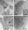
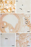
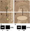
Methods and results: RT-PCR, sequencing and immunocytochemistry identified the genuine OTR in hGCs. OT (10 nM-10 microM) induced elevations of intracellular Ca(2+) levels (Fluo-4 measurements), which were blocked by tocinoic acid (TA; 50 microM, a selective OTR-antagonist). Down-stream effects of OTR-activation include a concentration dependent decrease in cell viability/metabolism, manifested by reduced ATP-levels, increased caspase3/7-activity (P < 0.05) and electron microscopical signs of cellular regression. TA blocked all of these changes. Immunoreactive OTR was found in the CL and GCs of large and, surprisingly, also small pre-antral follicles of the human ovary. Immunoreactive OTR in the rhesus monkey ovary was detected in primordial and growing primary follicles in the infantile ovary and in follicles at all stages of development in the adult ovary, as well as the CL: these results were corroborated by RT-PCR analysis of GCs excised by laser capture microdissection.
Conclusions: Our study identifies genuine OTRs in human and rhesus monkey GCs. Activation by high levels of OT leads to cellular regression in hGCs. As GCs of small follicles also express OTRs, OT may have as yet unknown functions in follicular development.
Figures


Similar articles
-
Decorin is a part of the ovarian extracellular matrix in primates and may act as a signaling molecule.Hum Reprod. 2012 Nov;27(11):3249-58. doi: 10.1093/humrep/des297. Epub 2012 Aug 11. Hum Reprod. 2012. PMID: 22888166 Free PMC article.
-
Progesterone receptor, but not estradiol receptor, messenger ribonucleic acid is expressed in luteinizing granulosa cells and the corpus luteum in rhesus monkeys.Endocrinology. 1994 Jul;135(1):307-14. doi: 10.1210/endo.135.1.8013365. Endocrinology. 1994. PMID: 8013365
-
Molecular cloning and functional characterization of the oxytocin receptor from a rat pancreatic cell line (RINm5F).Neuropeptides. 1996 Dec;30(6):557-65. doi: 10.1016/s0143-4179(96)90039-6. Neuropeptides. 1996. PMID: 9004255
-
Footmarks of innate immunity in the ovary and cytokeratin-positive cells as potential dendritic cells.Adv Anat Embryol Cell Biol. 2011;209:vii-99. doi: 10.1007/978-3-642-16077-6. Adv Anat Embryol Cell Biol. 2011. PMID: 21214088 Review.
-
Oxytocin/Oxytocin Receptor Signalling in the Gastrointestinal System: Mechanisms and Therapeutic Potential.Int J Mol Sci. 2024 Oct 11;25(20):10935. doi: 10.3390/ijms252010935. Int J Mol Sci. 2024. PMID: 39456718 Free PMC article. Review.
Cited by
-
The NADPH oxidase 4 is a major source of hydrogen peroxide in human granulosa-lutein and granulosa tumor cells.Sci Rep. 2019 Mar 5;9(1):3585. doi: 10.1038/s41598-019-40329-8. Sci Rep. 2019. PMID: 30837663 Free PMC article.
-
Human Luteinized Granulosa Cells-A Cellular Model for the Human Corpus Luteum.Front Endocrinol (Lausanne). 2019 Jul 9;10:452. doi: 10.3389/fendo.2019.00452. eCollection 2019. Front Endocrinol (Lausanne). 2019. PMID: 31338068 Free PMC article. Review.
-
Human tryptase cleaves pro-nerve growth factor (pro-NGF): hints of local, mast cell-dependent regulation of NGF/pro-NGF action.J Biol Chem. 2011 Sep 9;286(36):31707-13. doi: 10.1074/jbc.M111.233486. Epub 2011 Jul 18. J Biol Chem. 2011. PMID: 21768088 Free PMC article.
-
Progesterone inhibition of oxytocin signaling in endometrium.Front Neurosci. 2013 Aug 7;7:138. doi: 10.3389/fnins.2013.00138. eCollection 2013. Front Neurosci. 2013. PMID: 23966904 Free PMC article.
-
A Role for H2O2 and TRPM2 in the Induction of Cell Death: Studies in KGN Cells.Antioxidants (Basel). 2019 Oct 29;8(11):518. doi: 10.3390/antiox8110518. Antioxidants (Basel). 2019. PMID: 31671815 Free PMC article.
References
-
- Amico JA, Seif SM, Robinson AG. Oxytocin in human plasma: correlation with neurophysin and stimulation with estrogen. J Clin Endocrinol Metab. 1981a;52:988–993. - PubMed
-
- Amico JA, Seif SM, Robinson AG. Elevation of oxytocin and the oxytocin-associated neurophysin in the plasma of normal women during midcycle. J Clin Endocrinol Metab. 1981b;53:1229–1232. - PubMed
-
- Auletta FJ, Paradis DK, Wesley M, Duby RT. Oxytocin is luteolytic in the rhesus monkey (Macaca mulatta) J Reprod Fertil. 1984;72:401–406. - PubMed
-
- Bulling A, Berg FD, Berg U, Duffy DM, Stouffer RL, Ojeda SR, Gratzl M, Mayerhofer A. Identification of an ovarian voltage-activated Na+-channel type: hints to involvement in luteolysis. Mol Endocrinol. 2000;14:1064–1074. - PubMed
-
- Clayton B. Impairment of osmotically stimulated AVP release in patients with primary polydipsia. Am J Physiol. 1993;265:R1247. - PubMed
Publication types
MeSH terms
Substances
Grants and funding
LinkOut - more resources
Full Text Sources
Research Materials
Miscellaneous
