

Macular atrophy in birdshot retinochoroidopathy: an optical coherence tomography and multifocal electroretinography analysis
- PMID: 20098346
- PMCID: PMC2884075
- DOI: 10.1097/IAE.0b013e3181c720b4
Macular atrophy in birdshot retinochoroidopathy: an optical coherence tomography and multifocal electroretinography analysis
Abstract
Purpose: The purpose of this study was to evaluate macular atrophy by frequency-domain optical coherence tomography (OCT) in patients with birdshot retinochoroidopathy and to compare the resulting thickness measures with visual acuity and multifocal electroretinography (mfERG).
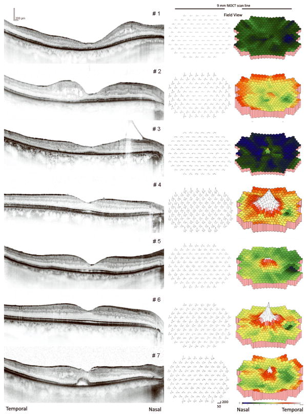
Methods: Measures were obtained from 14 eyes of 7 patients with birdshot retinochoroidopathy and 23 normal eyes. Optical coherence tomography-3 measures of macular thinning were related to visual acuity, mfERG response density, and time since diagnosis. Horizontal midline frequency-domain OCT scans identified which layers of the retina were primarily responsible for macular thinning.
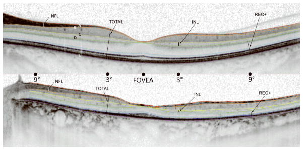
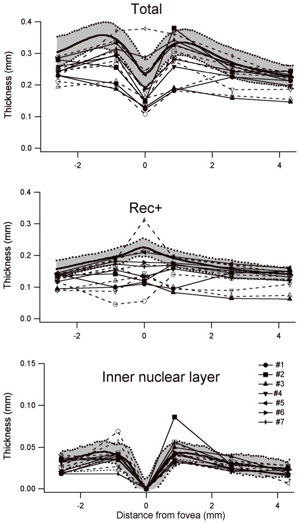
Results: All eyes with a history of birdshot retinochoroidopathy for >10 years had abnormal mfERG response densities. Compared with those without anatomic thinning (n = 8), eyes with anatomic thinning (n = 6) had significantly lower visual acuity (P = 0.0006), foveal response density (P = 0.006), and overall mfERG response density (P = 0.009). Segmentation of retinal layers on frequency-domain OCT scans showed that anatomic thinning was as a result of reduction in the receptor 1 layer (REC+), the thickness of the segment extending from the proximal border of the outer plexiform layer to the Bruch membrane-choroid interface.
Conclusion: Macular atrophy, as reflected in OCT evidence of macular thinning and mfERG evidence of macular function, occurs in patients with long-standing birdshot retinochoroidopathy. Measures of retinal layer thicknesses by frequency-domain OCT suggest that the atrophy occurs primarily in the outer retina.
Figures
Similar articles
-
Assessment of macular function in patients with idiopathic Epiretinal membrane by multifocal Electroretinography: correlation with visual acuity and optical coherence tomography.BMC Ophthalmol. 2017 Nov 28;17(1):221. doi: 10.1186/s12886-017-0621-1. BMC Ophthalmol. 2017. PMID: 29183292 Free PMC article.
-
Relation between macular retinal ganglion cell/inner plexiform layer thickness and multifocal electroretinogram measures in experimental glaucoma.Invest Ophthalmol Vis Sci. 2014 Jun 26;55(7):4512-24. doi: 10.1167/iovs.14-13937. Invest Ophthalmol Vis Sci. 2014. PMID: 24970256 Free PMC article.
-
Photoreceptor dysfunction in early and intermediate age-related macular degeneration assessed with mfERG and spectral domain OCT.Doc Ophthalmol. 2016 Feb;132(1):17-26. doi: 10.1007/s10633-016-9523-4. Epub 2016 Jan 11. Doc Ophthalmol. 2016. PMID: 26754967
-
[Pathophysiology of macular diseases--morphology and function].Nippon Ganka Gakkai Zasshi. 2011 Mar;115(3):238-74; discussion 275. Nippon Ganka Gakkai Zasshi. 2011. PMID: 21476310 Review. Japanese.
-
Microstructural and hemodynamic changes in the fundus after pars plana vitrectomy for different vitreoretinal diseases.Graefes Arch Clin Exp Ophthalmol. 2024 Jul;262(7):1977-1992. doi: 10.1007/s00417-023-06303-x. Epub 2023 Nov 20. Graefes Arch Clin Exp Ophthalmol. 2024. PMID: 37982887 Review.
Cited by
-
Multifocal electroretinograms.J Vis Exp. 2011 Dec 4;(58):3176. doi: 10.3791/3176. J Vis Exp. 2011. PMID: 22158462 Free PMC article.
-
Visual Field Tests: A Narrative Review of Different Perimetric Methods.J Clin Med. 2024 Apr 23;13(9):2458. doi: 10.3390/jcm13092458. J Clin Med. 2024. PMID: 38730989 Free PMC article. Review.
-
Phenotypic characterization of 3 families with autosomal dominant retinitis pigmentosa due to mutations in KLHL7.Arch Ophthalmol. 2011 Nov;129(11):1475-82. doi: 10.1001/archophthalmol.2011.307. Arch Ophthalmol. 2011. PMID: 22084217 Free PMC article.
-
Relationships among multifocal electroretinogram amplitude, visual field sensitivity, and SD-OCT receptor layer thicknesses in patients with retinitis pigmentosa.Invest Ophthalmol Vis Sci. 2012 Feb 21;53(2):833-40. doi: 10.1167/iovs.11-8410. Print 2012 Feb. Invest Ophthalmol Vis Sci. 2012. PMID: 22247460 Free PMC article.
-
An overview of optical coherence tomography angiography and the posterior pole.Ther Adv Ophthalmol. 2019 Apr 3;11:2515841419840249. doi: 10.1177/2515841419840249. eCollection 2019 Jan-Dec. Ther Adv Ophthalmol. 2019. PMID: 30984909 Free PMC article. Review.
References
-
- Ryan SJ, Maumenee AE. Birdshot retinochoroidopathy. Am J Ophthalmol. 1980;89:31–45. - PubMed
-
- Kaplan HJ, Aaberg TM. Birdshot retinochoroidopathy. Am J Ophthalmol. 1980;90:773–782. - PubMed
-
- Thorne JE, et al. Birdshot retinochoroidopathy: ocular complications and visual impairment. Am J Ophthalmol. 2005;140:45–51. - PubMed
-
- Gass JD. Vitiliginous chorioretinitis. Arch Ophthalmol. 1981:1778–1787. - PubMed
Publication types
MeSH terms
Grants and funding
LinkOut - more resources
Full Text Sources
Medical
