. 2010 Feb;12(2):111-8.
doi: 10.1038/ncb2011. Epub 2010 Jan 24.

DNA zip codes control an ancient mechanism for gene targeting to the nuclear periphery

Affiliations

DNA zip codes control an ancient mechanism for gene targeting to the nuclear periphery

Sara Ahmed et al. Nat Cell Biol. 2010 Feb.

Erratum in

  • Nat Cell Biol. 2010 Mar;12(3):306

Abstract

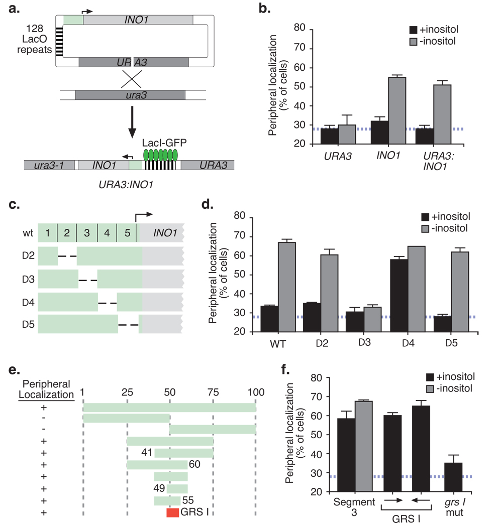
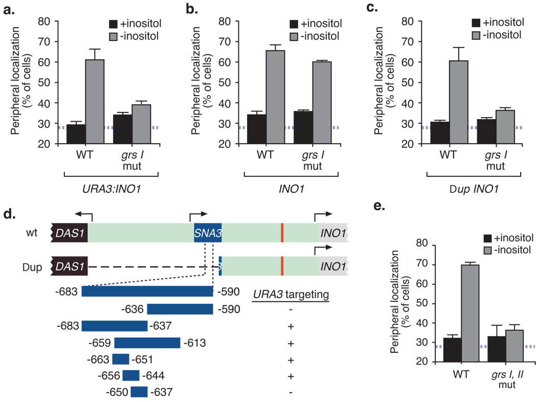
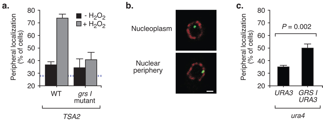
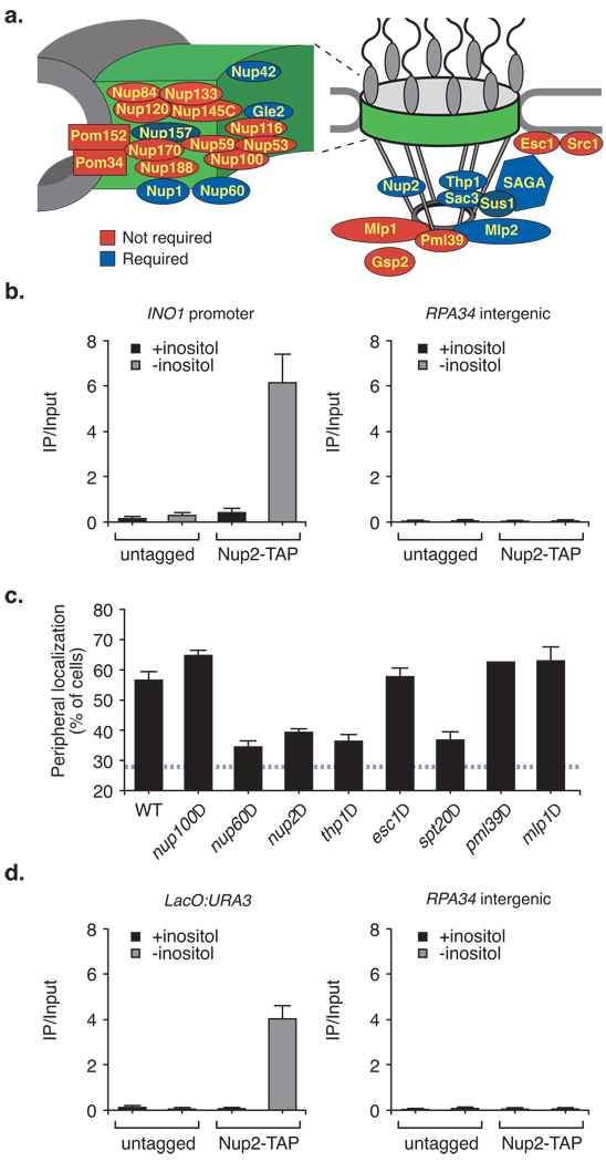
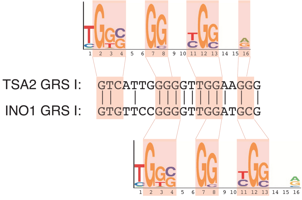
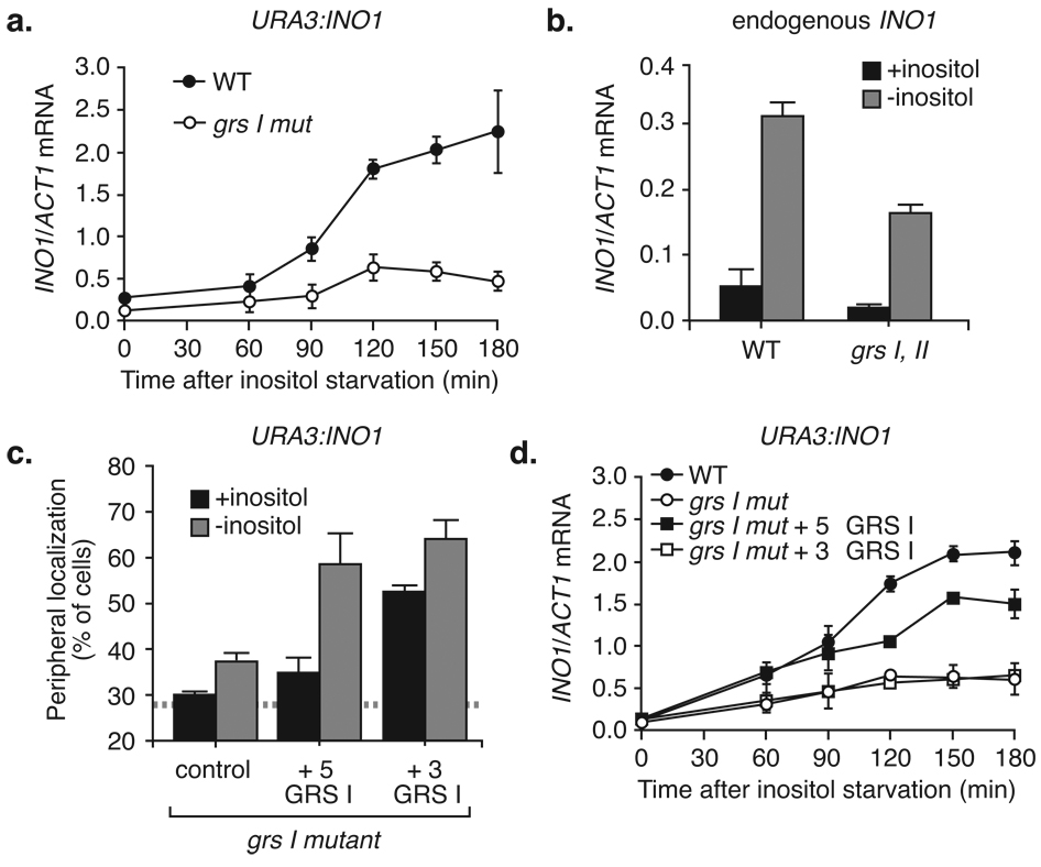
Many genes in Saccharomyces cerevisiae are recruited to the nuclear periphery after transcriptional activation. We have identified two gene recruitment sequences (GRS I and II) from the promoter of the INO1 gene that target the gene to the nuclear periphery. These GRSs function as DNA zip codes and are sufficient to target a nucleoplasmic locus to the nuclear periphery. Targeting requires components of the nuclear pore complex (NPC) and a GRS is sufficient to confer a physical interaction with the NPC. GRS I elements are enriched in promoters of genes that interact with the NPC, and genes that are induced by protein folding stress. Full transcriptional activation of INO1 and another GRS-containing gene requires GRS-mediated targeting of the promoter to the nuclear periphery. Finally, GRS I also functions as a DNA zip code in Schizosaccharomyces pombe, suggesting that this mechanism of targeting to the nuclear periphery has been conserved over approximately one billion years of evolution.


Figures

Figure 1
Figure 1. Identification of a Gene Recruitment Sequence (GRS) in the INO1 promoter
(a) Integration scheme for integrating INO1 and the lac repressor array at URA3 by homologous recombination. The INO1 gene included 504bp upstream and 685bp downstream of the coding sequence. (b) The fraction of the population in which the GFP spot colocalized with the nuclear envelope marker Sec63-myc for cells grown in the presence or absence of 100µM myo-inositol. The hatched blue line represents the mean peripheral localization for URA3. The maximal peripheral localization observed using this assay is ~80% of cells for a gene that is artificially tethered to the nuclear envelope. Therefore, the dynamic range of this assay is 20% – 80%. Data represent the mean and s.e.m from 5 biological replicates (30–50 cells were analyzed per replicate). Map (c) and peripheral localization (d) of 100bp non-overlapping deletions in the INO1 promoter integrated at URA3. (e) Map of fragments within segment 3 that were integrated at URA3 and their peripheral localization (complete data in Fig. S1a). (f) Peripheral localization of segment 3 in the presence and absence of inositol, the 8bp GRS I in either orientation or a mutant version of GRS I integrated at URA3 (n ≥ 3, 30–50 cells per biological replicate).
Figure 2
Figure 2. Targeting of the endogenous INO1 gene is mediated by two redundant DNA zip codes
(a) Localization of wild type INO1 and GRS I mutant INO1 integrated either at URA3 (a) or at INO1 (b). (c) Localization of wild type or grs I mutant INO1 at endogenous INO1 in a strain lacking the 943bp upstream of GRS I as depicted in panel (d) (for panels ad, n = 3, 30–50 cells per replicate). (d) Map of fragments used to identify GRS II by integration at URA3 and their peripheral localization (complete data in Figs. S1b and S1c). (e) Peripheral localization of a combined mutation in GRS I and GRS II at endogenous INO1 (n ≥ 3, 30–50 cells per replicate).
Figure 3
Figure 3. GRS I mediated targeting to the nuclear periphery is general and ancient
(a) Peripheral localization of the wild type or grs I mutant TSA2 gene was determined for a population of cells for cells grown in YPD (-H2O2 repressing conditions) or in YPD + 0.5mM H2O2 (activating conditions) (n = 3, 30–50 cells per replicate). (b) Introduction of the GRS into S. pombe. Representative confocal micrographs of immunofluorescence against LacI-GFP (green) and Nup120-myc (red) in S. pombe that were scored as either nucleoplasmic or peripheral. The scale bar equals 1µm. (c) The fraction of the population in which ura4 colocalized with Nup120-myc in cells with either the lac repressor array plasmid or the lac repressor array plasmid with a single copy of GRS I (n = 4, 60–100 cells per replicate).
Figure 4
Figure 4. INO1 recruitment to the nuclear periphery requires components of the nuclear pore complex (NPC) and associated factors
(a) Summary of nuclear pore proteins and associated factors required for INO1 targeting to the nuclear periphery (complete data in Fig. S3; data for Nup2 has previously been published12). Proteins filled in blue were required for INO1 peripheral targeting. Proteins filled in red were not required for INO1 peripheral targeting. The positions of proteins within the pore and contacts between them are approximations based on a structural model of the NPC structure. Left: an expanded view of the core channel of the NPC. (b) Chromatin immunoprecipitation of Nup2-TAP under repressing and activating conditions. Recovery of the INO1 promoter (left panel) or the RPA34 intergenic region ~5000 bp upstream (right panel) with IgG magnetic beads (Invitrogen) was quantified relative to the input by real-time quantitative PCR. (c) Nuclear pore requirements for GRS I mediated targeting of URA3 (n ≥ 2, 30–50 cells per replicate). Cells were grown in the presence of inositol. (d) Recovery of URA3 or RPA34 with Nup2 (as in panel b) from strains with or without GRS I at the URA3 locus (see Methods). For panels b and d, data represent the mean and the s.e.m. (n = 3).
Figure 5
Figure 5. GRS I is enriched among genes that interact with many nuclear pore proteins
Comparison of GRS I from INO1 and TSA2 to a motif, identified by Casolari et al., that is overrepresented in genes associated with Mlp1, Mlp2 or Nic96. Two alternative alignments are shown.
Figure 6
Figure 6. Localization at the nuclear periphery enhances transcription of INO1
(a) INO1 mRNA levels were quantified by RT-qPCR, relative to ACT1, following induction by inositol starvation from strains having either plasmid-borne wild type INO1 or grs I mutant INO1 integrated at URA3. (b) Strains were constructed in which the grs I, II mutations were introduced at the chromosomal INO1 locus. Wild type and grs I, II mutant strains were grown overnight in the presence or absence of inositol and the mRNA levels quantified as in (a). (c & d) GRS I was reintroduced either at the 5’ end or the 3’ end of grs I mutant INO1 and these plasmids were integrated at URA3. These strains were compared with wild type and grs I mutant INO1 for localization (c) and transcription (d). For panels ad, n = 4.

Similar articles

Cited by

References

    1. Akhtar A, Gasser SM. The nuclear envelope and transcriptional control. Nat Rev Genet. 2007;8:507–517. - PubMed
    1. Ahmed S, Brickner JH. Regulation and epigenetic control of transcription at the nuclear periphery. Trends Genet. 2007;23:396–402. - PubMed
    1. Taddei A. Active genes at the nuclear pore complex. Curr Opin Cell Biol. 2007;19:305–310. - PubMed
    1. Casolari JM, et al. Genome-wide localization of the nuclear transport machinery couples transcriptional status and nuclear organization. Cell. 2004;117:427–439. - PubMed
    1. Brickner JH, Walter P. Gene recruitment of the activated INO1 locus to the nuclear membrane. PLoS Biol. 2004;2:e342. - PMC - PubMed

Publication types

MeSH terms

Substances