Skin dose estimation for various beam modifiers and source-to-surface distances for 6MV photons
- PMID: 20098542
- PMCID: PMC2805895
- DOI: 10.4103/0971-6203.51935
Skin dose estimation for various beam modifiers and source-to-surface distances for 6MV photons
Abstract
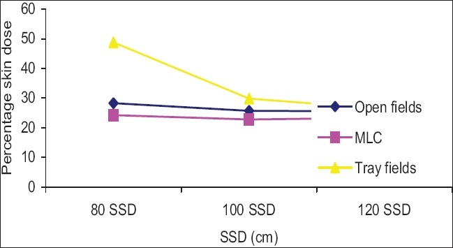
The purpose of this study was to learn the skin dose estimation for various beam modifiers at various source-to-surface distances (SSDs) for a 6 MV photon. Surface and buildup region doses were measured with an acrylic slab phantom and Markus 0.055 cc parallel plate (PP) ionization chamber. Measurements were carried out for open fields, motorized wedge fields, acrylic block tray fields ranging from 3 x 3 cm(2) to 30 x 30 cm(2). Twenty-five percent of the field was blocked with a cerrobend block and a Multileaf collimator (MLC). The effect of the blocks on the skin dose was measured for a 20 x 20 cm(2) field size, at 80 cm, 100 cm and 120 cm SSD. During the use of isocentric treatments, whereby the tumor is positioned at 100 cm from the source, depending on the depth of the tumor and size of the patient, the SSD can vary from 80 cm to 100 cm. To achieve a larger field size, the SSD can also be extended up to 120 cm at times. The skin dose increased as field size increased. The skin dose for the open 10 x10 cm(2) field was 15.5%, 14.8% and 15.5% at 80 cm, 100 cm and 120 cm SSDs, respectively. The skin dose due to a motorized 60 degrees wedge for the 10 x 10 cm(2) field was 9.9%, 9.5%, and 9.5% at 80 cm, 100 cm and 120 cm SSDs. The skin dose due to acrylic block tray, of thickness 1.0 cm for a 10 x 10 cm(2) field was 27.0%, 17.2% and 16.1% at 80, 100 and 120 cm SSD respectively. Due to the use of an acrylic block tray, the surface dose was increased for all field sizes at the above three SSDs and the percentage skin dose was more dominant at the lower SSD and larger field size. The skin dose for a 30 x 30 cm(2) field size at 80 cm SSD was 38.3% and it was 70.4% for the open and acrylic block tray fields, respectively. The skin doses for motorized wedge fields were lower than for open fields. The effect of SSDs on the surface dose for motorized 60 degrees wedge fields was not significant for a small field size (difference was less than 1% up to a 15 x 15 cm(2) field size), but for a larger field (field size more than 15 x 15 cm(2)), the difference in a percentage skin dose was significant. The skin dose for the open field was more than that for the MLC blocked field and lower than that for the acrylic blocked tray field. The block was 25% of the 20 x 20 cm(2) open field. Skin doses were increased as the SSD decreased and were dominant for larger field sizes. The surface dose was weakly dependent on the MLC block.
Keywords: Acrylic block Tray; Block; Field size; Motorized 60° wedge; Multileaf collimator; Percentage skin dose; Surface dose; source-to-surface distance.
Conflict of interest statement
Figures


References
-
- Sixel K, Podgorsak E. Buildup region and depth of dose maximum of mega voltage X – ray beams. Med Physics. 1994;21:411–6. - PubMed
-
- Kim S, Liu CR, Zhu TC, Palta JR. Photon beam skin dose analysis for different clinical setups. Med Phys. 1998;25:860–6. - PubMed
-
- Nizin PZ. Electronic equilibrium and primary dose in collimated photon beams. Med Phys. 1993;20:1721–9. - PubMed
-
- Pettti PL, Goodman MS, Sisterson JM. Sources of electron contamination for the clinc-35, 25 –MV- photon beam. Med Phys. 1983;10:856–61. - PubMed
-
- Nilsson B, Brahme A. Electron contamination from photon beam collimators. Radiother Oncol. 1986;5:235–44. - PubMed
LinkOut - more resources
Full Text Sources
Miscellaneous