. 2008 Apr 5:3:91.
doi: 10.1007/7050_2008_039.

Biochemistry of Meiotic Recombination: Formation, Processing, and Resolution of Recombination Intermediates

Affiliations

Biochemistry of Meiotic Recombination: Formation, Processing, and Resolution of Recombination Intermediates

Kirk T Ehmsen et al. Genome Dyn Stab. .

Abstract

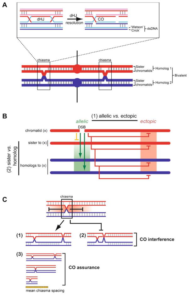
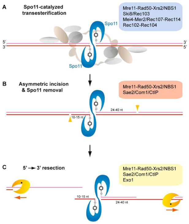
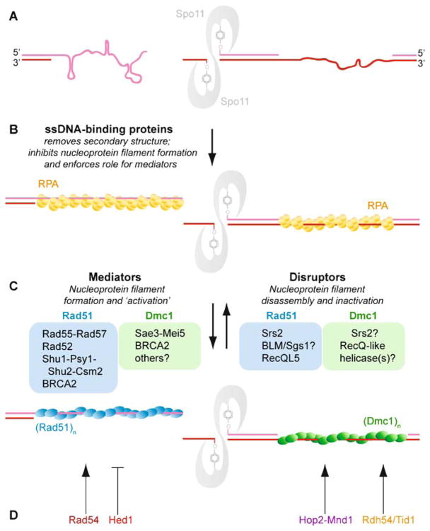
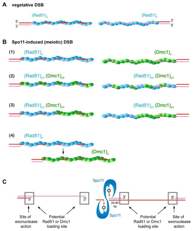
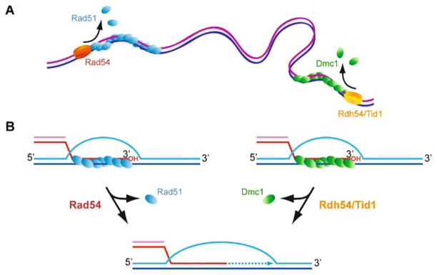
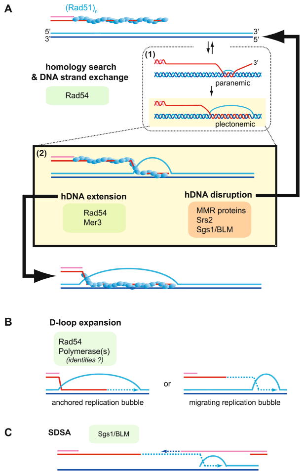
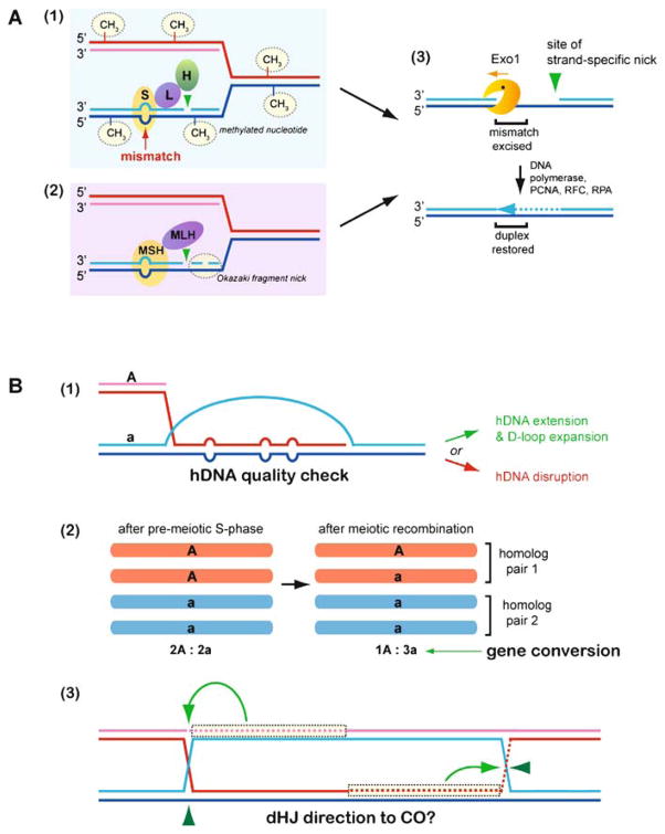
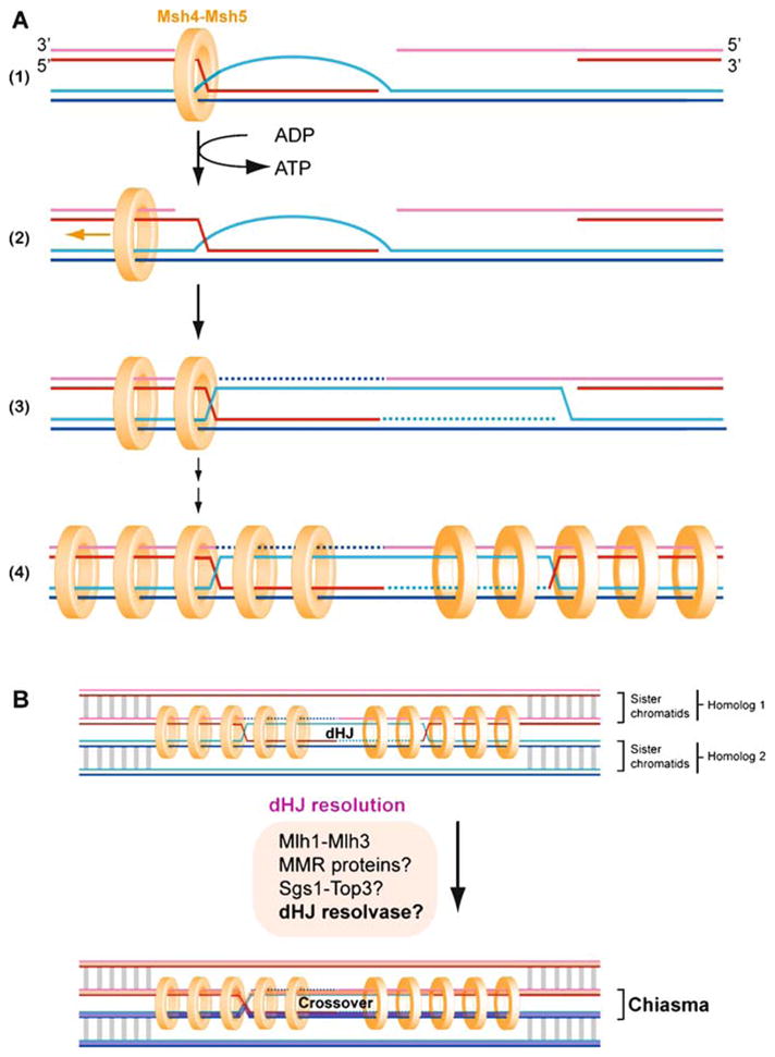
Meiotic recombination ensures accurate chromosome segregation during the first meiotic division and provides a mechanism to increase genetic heterogeneity among the meiotic products. Unlike homologous recombination in somatic (vegetative) cells, where sister chromatid interactions prevail and crossover formation is avoided, meiotic recombination is targeted to involve homologs, resulting in crossovers to connect the homologs before anaphase of the first meiotic division. The mechanisms responsible for homolog choice and crossover control are poorly understood, but likely involve meiosis-specific recombination proteins, as well as meiosis-specific chromosome organization and architecture. Much progress has been made to identify and biochemically characterize many of the proteins acting during meiotic recombination. This review will focus on the proteins that generate and process heteroduplex DNA, as well as those that process DNA junctions during meiotic recombination, with particular attention to how recombination activities promote crossover resolution between homologs.


Figures

Fig. 1
Fig. 1
Meiotic crossovers establish physical connection between homologs. A The two homologs each consist of two sister chromatids (red and blue lines each depicting a dsDNA molecule) held together by cohesion (represented by grey lines between the sisters). Upon bipolar attachment of the kinetochores (red/blue circles) to the meiosis I spindle (black lines), the CO points between homologs (indicated as chiasmata) provide a counterforce to the spindle force acting on the kinetochores, signaling correct bipolar attachment of the paired homologs (bivalent) and ensuring high-fidelity chromosome segregation during meiosis I division. Resolution of a double-Holliday junction (dHJ) by alternate incision, as shown in the box, represents a mechanism to generate a chiasma. The individual DNA strands involved are shown in the box. B Meiotic recombination entails objectives unique from those in vegetative cells. (1) As in vegetative cells, recombination is minimized between ectopic sequences but is instead directed toward allelic sites. Mechanisms responsible for the biochemical differentiation between ectopic (homeologous) and allelic (homologous) sites are poorly known but probably involve mismatch repair factors and regulation at the level of heteroduplex quality during preliminary DNA strand exchange events. (2) Meiotic recombination promotes a regulated level of DSB repair directed to the homolog, at the exclusion of the sister chromatid. The biochemical basis of sister vs. homolog discrimination is also poorly understood and remains an outstanding question for recombination applications specific to meiosis. C Crossovers are an essential outcome of the meiotic recombination agenda, but only under strict limitations of number (incidence) and distribution. Where one CO occurs in a bivalent, the probability of a second CO nearby is far below what would be expected by random distribution. This suggests that the number and spacing of COs is regulated; a phenomenon known as CO (or chiasma) interference. It is unclear at what level (pre-DSB, DSB, SEI, dHJ) interference is imposed. Not all organisms display interference (e.g., Schizosaccharomyces pombe does not) (Munz 1994), and not all CO pathways are associated with interference (see Fig. 12). Although the underlying mechanism(s) for CO interference remain to be explained, interference results in the non-random spacing of chiasmata on chromosomes that undertake multiple CO events (1) and (2). Interference may also play a role in CO assurance (3), the observation that all bivalents earn at least one chiasma, even on chromosomes that are smaller than the mean chiasma spacing
Fig. 2
Fig. 2
Mechanistic stages of homologous recombination. Meiotic recombination is initiated by a Spo11-mediated double-stranded DNA break (DSB) (1). During presynapsis, the initial break is resected to form 3′-OH ending single-stranded DNA tails to allow formation of filaments by the DNA strand exchange proteins, Rad51 and Dmc1 (2). During synapsis, a joint molecule is formed between the broken DNA and an unbroken template from the other homolog, positioning the 3′-OH end for DNA synthesis (3). During postsynapsis, meiotic recombination bifurcates into at least two primary pathways that repair the DSBs: most breaks are repaired to NCO products by SDSA, but a fraction of breaks are repaired to CO products by DSBR. SDSA (4b, 5b) dissolves the initial D-loop to reanneal the extended invading strand to the second end of the break site, resulting in NCO products (Nassif et al. 1994; Resnick 1976). Second end capture and dHJ formation (DSBR, 5a, 6a,b, 7) (Szostak et al. 1983) account for the main CO pathway in budding yeast, nematodes, and mammals (termed CO pathway 1). Possible scenarios for CO pathway 2 (predominant in fission yeast) and 3 (predominant in Drosophila) are shown in Fig. 12. The joint molecule physically identified as the SEI intermediate (4a) appears to be a stabilized D-loop and is a CO-specific intermediate in meiosis (Hunter and Kleckner 2001). The dHJ intermediate (5a) is critical for CO formation, possibly through resolution by structure-specific endonucleases resembling the bacterial RuvC enzyme (6a). Resolution of dHJs might be biased to CO products, such that there is no NCO outcome (“?” in 5a to 5b transition). Alternatively, a minor fraction of dHJs may be dissolved into NCO products by a RecQ-family helicase, a topoisomerase III, and a junction specificity factor, involving reverse-branch migration that confines heteroduplex DNA to the recipient chromosome (6b-7) (Wu and Hickson 2003) (see Fig. 11)
Fig. 3
Fig. 3
Spo11-catalyzed DSBs and asymmetric end processing by 5′ → 3′ resection. A Spo11 is a type II-related topoisomerase that catalyzes DSB formation at “hotspots” by a transesterification mechanism involving a covalent intermediate between a tyrosine residue of a Spo11 subunit and each 5′ end. A legion of factors (see blue box) is implicated in Spo11 DSB initiation, and their biochemical contributions to Spo11 activity need explanation. B Spo11 remains covalently bound to its product DNA break ends and can be isolated in two populations, bound to oligonucleotides of asymmetric lengths (Neale et al. 2005). The enzymes involved in the endonucleotlytic incision are shown in the red box. One population is associated with short oligonucleotides, 10–15 nt, while a second population is recovered in association with oligonucleotides 24–40 nt in length. This result provides a possible mechanism to establish asymmetry of break ends at or near the timing of Spo11-catalyzed DSBs, although the underlying basis of the asymmetry is unknown. The two populations of oligonucleotide-Spo11 complexes imply that 5′ → 3′ resection initiates at nicks positioned asymmetrically to the Spo11 cleavage complex. C A number of factors are implicated in DNA end resection (see yellow box), but the primary exonuclease or endonuclease activities remain uncertain. Resection is processive up to ~ 500 nt on each break end and is possibly coupled to Rad51 and Dmc1 loading. The short oligonucleotide-Spo11 complex (10–15nt) is suggested to separate readily from its complementary strand, generating a free 3′ end (Neale et al. 2005). The longer oligonucleotide-Spo11 complex (24–40nt) may remain paired to its complementary strand and therefore resection may generate a gapped region instead of a free end. These asymmetries may imply differential assembly of Rad51 and Dmc1 filaments (see Fig. 5). Alternatively, the oligonucleotides associated with Spo11 may remain base-paired to their complements, but the larger duplex extent on one side may present a binding site or interaction surface for a mediator protein specific for Dmc1 or Rad51 (see Fig. 4)
Fig. 4
Fig. 4
Regulation of Rad51 and Dmc1 nucleoprotein filament formation and function. A The ssDNA generated by 5′ → 3′ resection is likely to form secondary structures inhibitory to the formation of active Rad51 or Dmc1 nucleoprotein filaments. B RPA overcomes this challenge to Rad51 or Dmc1 by melting ssDNA secondary structure during binding, but RPA binds avidly to ssDNA and therefore inhibits nucleoprotein filament formation because RPA is poorly displaced by Rad51 or Dmc1 alone. C Aside from indirectly promoting nucleoprotein filament formation on ssDNA, RPA presents an opportunity for regulation of Rad51 or Dmc1 loading on ssDNA by enforcing a role for mediators. Mediators are a class of proteins that can best be characterized as factors that promote functional filaments (assayed by capacity for DNA strand exchange), although the mechanisms by which they promote a functional filament may be diverse and include: (1) regulation of RPA displacement and Rad51 nucleation on ssDNA, (2) regulation of Rad51 stability (turnover rates) on ssDNA, at an dsDNA-ssDNA junction, or on heteroduplex DNA, (3) filament nucleation and regulation of filament growth, or (4) a function with the free subunit pool. The asymmetry of Spo11 cleavage complexes may suggest a role for mediators specific to nucleation of Rad51 or Dmc1 at one or the other face of the cleavage complex. In some contexts, Srs2 may be considered a mediator of functional filaments if its displacement of Rad51 allows proper registry of a contiguous filament rather than small Rad51 patches on a ssDNA lattice that maybe out of register from one another. Biochemically, Srs2 removes Rad51 from ssDNA; Dmc1 remains to be tested. D Other factors further regulate the function of an assembled Rad51-ssDNA or Dmc1-ssDNA filament. Rad54 stabilizes the Rad51-ssDNA filament and promotes the DNA strand exchange activity of Rad51 filaments by mechanisms that may include topological remodeling of the dsDNA target. Rdh54/Tid1 also stimulates Rad51 DNA strand exchange (Petukhova et al. 2000), and by analogy, Rdh54/Tid1 likely interacts with the Dmc1 filament to promote its DNA strand exchange activity. Genetically, Hed1 appears to inhibit DNA strand exchange activity catalyzed by the Rad51 filament, but biochemical data is lacking. Hop2-Mnd1 promotes duplex capture by the Dmc1 filament
Fig. 5
Fig. 5
Models for Dmc1 and Rad51-induced DSB ends: cofilaments or asymmetric filaments. A Rad51 is the sole RecA homolog employed during vegetative recombination in S. cerevisiae. It assembles as symmetric filaments on each DSB end, although each end may be differentially regulated (for DNA strand exchange or second-end capture in DSBR) and these details await biochemical explanation. B Dmc1 is a meiosis-specific Rad51 paralog that functions in collaboration with Rad51 for the purposes of homolog-directed DNA strand exchange with resolution to CO. There are at least four possibilities for the collaborative relationship of Rad1 and Dmc1 in filaments: 1 Rad51 and Dmc1 may assemble as separate filaments on each break end; 2 Rad51 and Dmc1 may assemble as mixed filaments on each break end; 3 Rad51 and Dmc1 may assemble as patchy cofilaments on each break end; or 4 Rad51 and Dmc1 may assemble consecutively on the same ssDNA regions during different stages of meiotic recombination. C The asymmetry suggested for Spo11-induced DSB processing (Neale et al. 2005) presents several opportunities to direct different loading of Rad51 and Dmc1 to one or the other break end. Furthermore, the break ends may remain associated but the different oligo lengths adjacent to the Spo11 homodimer may present binding sites for recruitment of Rad51- or Dmc1-specific mediators
Fig. 6
Fig. 6
Rad54 and Rdh54-Tid1: removal of Rad51 from dsDNA and DNA polymerase extension from the 3′ end of heteroduplex DNA. Rad51 and Dmc1 bind readily to dsDNA, unlike their bacterial counterpart RecA. Rad54 and Rdh54/Tid1 may function to dissociate Rad51-dsDNA complexes or Dmc1-dsDNA complexes, in at least two contexts: A non-specific binding to dsDNA (“dead-end” complexes), and B turnover from the heteroduplex dsDNA product of DNA strand exchange to allow access of DNA polymerase to the invading 3′ end. Not all activities depicted here have been experimentally demonstrated, but are inferred from in vivo and in vitro results with these proteins (for details see text)
Fig. 7
Fig. 7
Joint molecule dissociation versus maturation by hDNA extension and D-loop expansion. A Homology search and DNA strand invasion by the Rad51 (or Dmc1, not shown) filament leads to the D-loop intermediate. Strand invasion by an incomplete filament or DNA strand invasion initiating internally (not at end) generates a paranemic joint, where the invading strand is not fully intertwined with the template strand and pairing is protein-mediated. (See box 1 for a representation, although the true nature of a paranemic interaction is uncertain. The Rad51 protein is not shown for simplicity.) Paranemic joints are unstable and may revert, or the pairing is extended to the end, allowing formation of a plectonemic joint with full strand intertwining in which heteroduplex base-pairing is sufficient for stability of the DNA strand exchange product (as explicitly drawn in box 1). Rad51 filament nucleation at the dsDNA–ssDNA junction would increase the probability of interstitial pairing resulting in paranemic joints. The D-loop drawn in box 2 is a plectonemic joint, but for simplicity strand intertwining is not drawn. The D-loop may be disrupted in processes averting recombination or during SDSA (after DNA synthesis, see C) involving MMR proteins, Srs2, and RecQ-like DNA helicases (see text for details). Alternatively, the D-loop can be enlarged by hDNA extension, and the Rad54 motor protein as well as the Mer3 DNA helicase have been implicated in this step. Mer3 plays a key role in CO formation through CO pathway 1 (see text). The extended D-loop is possibly the metastable intermediate SEI that is specific for CO pathway 1 (Hunter and Kleckner 2001). B The initial D-loop may also be expanded by DNA synthesis as an anchored bubble (left) or may become a migrating bubble (right), where its size remains unchanged (Formosa and Alberts 1986). C SDSA is effectively D-loop expansion coupled to regulated hDNA and D-loop disruption. Presumably, D-loop disruption is initiated after homology quality check has sanctioned DNA synthesis. hDNA extension and D-loop expansion are not necessarily stable end-points. DNA strand invasion and SDSA may be dynamic sampling states, where joint molecule formation and disruption occur iteratively, explaining the identification of genetic information obtained from multiple donor sites (Symington and Heyer 2006)
Fig. 8
Fig. 8
Applications of mismatch repair to meiotic recombination. A Signals in the parental versus daughter DNA strands distinguish a newly replicated DNA strand from its template strand, and are used by mismatch repair proteins to target excision repair to the new strand and its misincorporated nucleotide. (1) Prokaryotic mismatch repair capitalizes on the hemimethylated state of newly replicated DNA; MutS (indicated as S) scans the DNA for discontinuities, to which it binds and in association with MutL (indicated as L), presents to the endonuclease MutH (indicated as H). (2) Eukaryotic mismatch repair, involving MutS and MutL homologs (MSH, MHL), identifies the newly replicated strand not based on an absent signal (absence of methylation), but more likely on the presence of a nick associated with Okazaki fragment processing during lagging strand DNA synthesis. As in prokaryotic MMR, the nick defines the starting point for an exonuclease that travels to the mismatched region and removes the misincorporated nucleotide(s). (3) DNA repair synthesis and ligation restores the helix. B The basic properties of mismatch repair (mismatch binding, structural recognition, DNA strand discrimination, and endonuclease instruction) maybe employed for specific objectives of meiotic recombination. Several possibilities are shown here. (1) MMR factors bound to mismatches within heteroduplex DNA likely report on the degree of homeology or homology, which may distinguish ectopic from allelic targets. How MMR factors engage with a Rad51 or Dmc1 filament on hDNA is unknown and how they communicate to sanction hDNA extension or promote hDNA disruption is also unknown. (2) Repair of mismatches by MMR pathways is responsible for gene conversion. (3) The dHJ intermediate in budding yeast contains contiguous strands (Schwacha and Kleckner 1995), but a prior nick or another feature associated with DNA repair synthesis in CO pathway 1 may inform the placement of strand incisions by a dHJ resolvase, enforcing a CO outcome without a need for the two resolution events to be directly coordinated
Fig. 9
Fig. 9
Msh4-Msh5 promotes crossovers by stabilizing joint molecules associated with single-end invasions. A Msh4-Msh4 loads at a joint (single-end invasion). An ADP→ATP switch induces Msh4-Msh5 to slide on duplex DNA, embracing both homologs within a ring-like complex. Iterative loading of Msh4-Msh5 complexes and their subsequent diffusion is thought to tether the homologs (Snowden et al. 2004). Loading at only one joint is shown, and it is unclear whether Msh4-Msh5 loads at the invasion intermediate or at both nicked Holliday junctions. Ligation of the nicked junctions results in covalently sealed dHJs, the intermediates presently believed to precede resolution to CO outcome in CO pathway 1 (Schwacha and Kleckner 1995). In association with Mlh1-Mlh3, MMR proteins, Sgs1-Top3, and/or an unidentified dHJ resolvase, dHJ resolution is directed or enforced to CO. B The Msh4-Msh5 dHJ and CO resolution to a chiasma is shown in the context of the bivalent; cohesion is depicted by gray lines and may also be rings encircling the chromatids rather than the homologs as by Msh4-Msh5. The loss of cohesion associated with Msh4-Msh5 loading on homologs may partially account for CO interference in CO pathway 1 (see Fig. 1), as loss of cohesion over long tracks associated with closely spaced Msh4-Msh5 COs may risk premature loss of sister chromatid cohesion
Fig. 10
Fig. 10
XPF-ERCC1 promotes long-tract gene conversion by loop incision in a manner different from its incision during nucleotide incision repair. A XPF-ERCC1 is biochemically responsible for “upper-strand incision” 5′ to a single-stranded DNA lesion during nucleotide excision repair (NER). RPA promotes cleavage of the bubble substrate. B In contrast to its behavior in NER, XPF-ERCC1 is proposed to incise the “bottom strand” of a looped substrate associated with a long-tract heterology during gene conversion (Jensen et al. 2005). This incision site is genetically implicated but remains to be biochemically demonstrated in association with the factors that might direct or promote its incision in this context (such as Msh2-Msh6). C The predicted incision site for XPF-ERCC1 in long-tract gene conversion promotes the preservation of long insertions in otherwise homologous sequences
Fig. 11
Fig. 11
Double-Holliday junction resolution and dissolution: CO and NCO outcomes. A dHJ resolution: dHJ incision by a DNA structure-specific endonuclease can yield CO or NCO outcome, depending on whether the same strands or different strands are cut at each junction. dHJs are recognized as intermediates of CO pathway 1 and are thought to be resolved primarily, if not exclusively, to CO outcome. Inset: HJ in open planar configuration, showing symmetric incision across its core. Yellow bubbles represent the two phospho-diester bonds hydrolyzed during HJ resolution. B dHJ dissolution: dHJ can be dissolved to NCO outcome by coordinated convergent migration of the Holliday junctions followed by their decatenation, a mechanism demonstrated for BLM-TOPOIIIa-BLAP75. Inset: Single-strand DNA passage at a hemicatenane (collapsed dHJs) accounts for the separation to NCO. The yellow bubble represents the single phospho-diester bond hydrolyzed during DNA strand passage. Although the phospho-diester bond hydrolysis penultimate to strand passage probably occurs once at the single hemicatenane, topoisomerase nicking likely occurs repeatedly during dHJ convergent migration to relieve torsional strain associated with branch migration
Fig. 12
Fig. 12
Pathways to generate crossovers. A Symmetric endonucleolytic resolution of dHJs (a) (Szostak et al. 1983) and single HJs b′ (Holliday 1964) have long been considered to represent two mechanisms of generating crossovers. dHJ resolution represents CO pathway 1. CO pathway 2 is currently defined by Mus81-Mms4/Eme1, although the physical intermediates of this pathway are presently uncertain. A single HJ can be generated by endonucleolytic cleavage 3′ to an extended D-loop (b), if the displaced strand of the D-loop reanneals with the second end b′. Two successive rounds of D-loop cleavage, or more specifically D-loop incision at the bottom strand (c) and nicked HJ incision c′, can generate a CO without the involvement of an intact HJ intermediate (Heyer et al. 2003; Osman et al. 2003). Finally, consecutive incision of nicked dHJs (d) can yield a CO product. B Taxonomic distribution of CO pathways. CO pathway 1 is defined by a collection of factors associated with Msh4-Msh5, including Mer3, Mlh1-Mlh3, and synaptonemal complex factors not discussed here. CO pathway 2 is defined by the XPF paralog Mus81-Mms4/Eme1, and CO pathway 3 by the XPF ortholog Mei9 (pathway 3). The size of the checked box indicates the relative use of the pathway in the given organism. (*) Mus81-Mms4/Eme1 and Mei9-Mus312 are the endonucleases responsible for the incisions believed to be associated with CO generation in CO pathways 2 and 3, but Msh4-Msh5 is not an endonuclease and the endonuclease responsible for dHJ resolution in CO pathway 1 remains to be identified. CO pathway 1 displays CO interference (see Fig. 1). Interference is not intrinsic to chiasmata, as COs in pathway 2 do not display interference (de los Santos et al. 2003; Munz 1994). In Drosophila melanogaster, most if not all COs depend on pathway 3 (Sekelsky et al. 1995), implying that COs in this pathway are associated with interference (Muller 1916; Sturtevant 1915)

Similar articles

Cited by

References

    1. Aboussekhra A, Chanet R, Adjiri A, Fabre F. Semi-dominant suppressors of Srs2 helicase mutations of Saccharomyces cerevisiae map in the RAD51 gene, whose sequence predicts a protein with similarities to procaryotic RecA protein. Mol Cell Biol. 1992;12:3224–3234. - PMC - PubMed
    1. Adams MD, McVey M, Sekelsky JJ. Drosophila BLM in double-strand break repair by synthesis-dependent strand annealing. Science. 2003;299:265–267. - PubMed
    1. Akamatsu Y, Dziadkowiec D, Ikeguchi M, Shinagawa H, Iwasaki H. Two different Swi5-containing protein complexes are involved in mating-type switching and recombination repair in fission yeast. Proc Natl Acad Sci USA. 2003;100:15770–15775. - PMC - PubMed
    1. Akamatsu Y, et al. Fission yeast Swi5/Sfr1 and Rhp55/Rhp57 differentially regulate Rhp51-dependent recombination outcomes. EMBO J. 2007;26:1352–1362. - PMC - PubMed
    1. Alani E, Padmore R, Kleckner N. Analysis of wild-type and rad50 mutants of yeast suggest an intimate relationship between meiotic chromosome synapsis and recombination. Cell. 1990;61:419–436. - PubMed

LinkOut - more resources