. 2010 Jan 20;5(1):e8780.
doi: 10.1371/journal.pone.0008780.

GABA increases electrical excitability in a subset of human unmyelinated peripheral axons

Affiliations

GABA increases electrical excitability in a subset of human unmyelinated peripheral axons

Richard W Carr et al. PLoS One. .

Abstract

Background: A proportion of small diameter primary sensory neurones innervating human skin are chemosensitive. They respond in a receptor dependent manner to chemical mediators of inflammation as well as naturally occurring algogens, thermogens and pruritogens. The neurotransmitter GABA is interesting in this respect because in animal models of neuropathic pain GABA pre-synaptically regulates nociceptive input to the spinal cord. However, the effect of GABA on human peripheral unmyelinated axons has not been established.

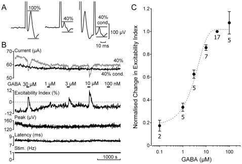
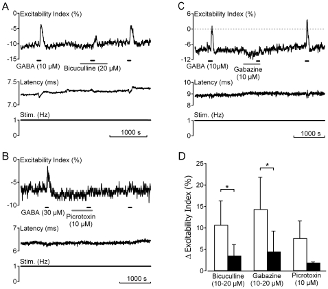
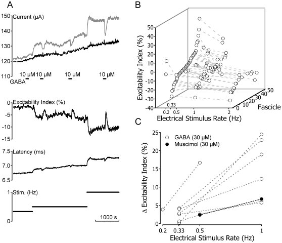
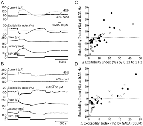
Methodology/principal findings: Electrical stimulation was used to assess the effect of GABA on the electrical excitability of unmyelinated axons in isolated fascicles of human sural nerve. GABA (0.1-100 microM) increased electrical excitability in a subset (ca. 40%) of C-fibres in human sural nerve fascicles suggesting that axonal GABA sensitivity is selectively restricted to a sub-population of human unmyelinated axons. The effects of GABA were mediated by GABA(A) receptors, being mimicked by bath application of the GABA(A) agonist muscimol (0.1-30 microM) while the GABA(B) agonist baclofen (10-30 microM) was without effect. Increases in excitability produced by GABA (10-30 microM) were blocked by the GABA(A) antagonists gabazine (10-20 microM), bicuculline (10-20 microM) and picrotoxin (10-20 microM).

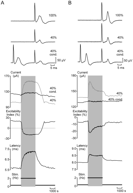
Conclusions/significance: Functional GABA(A) receptors are present on a subset of unmyelinated primary afferents in humans and their activation depolarizes these axons, an effect likely due to an elevated intra-axonal chloride concentration. GABA(A) receptor modulation may therefore regulate segmental and peripheral components of nociception.


Conflict of interest statement

Competing Interests: The authors have declared that no competing interests exist.

Figures

Figure 1
Figure 1. GABA activation of human C-fibres is concentration dependent.
The excitability index was determined for C-fibres in human sural nerve fascicles during bath application of GABA (0.1–100 µM). A time window restricted the domain over which the amplitude of the electrically-evoked compound C-fibre action potential was determined (A, grey bars). Excitability index was calculated from the ratio of the current required to evoke an unconditioned C-fibre response of 40% maximum amplitude to that required to evoke a conditioned 40% response, i.e. 30 ms after a supra-maximal conditioning stimulus (40% cond., grey). Negative values of excitability index indicate that more current is required to evoke an unconditioned 40% C-fibre response. Following the addition of GABA (0.1–30 µM, 90 s application) to the bathing solution the excitability index increases, i.e. becomes more positive (B), and the magnitude of this change increases as the concentration of GABA in the perfusing solution increases (B & C). The EC50 determined from a sigmoid fit to normalised excitability index on GABA concentration was 6.88±0.01 µM.
Figure 2
Figure 2. GABAA receptors mediate responses to GABA in human C-fibres.
Increases in the electrical excitability index of unmyelinated axons in human sural nerve fascicles following bath application of GABA (10–30 µM) are blocked by prior application of the GABAA receptor antagonists bicuculline (20 µM, grey bar A), picrotoxin (10 µM, grey bar B) and gabazine (10 µM, grey bar C). In contrast to both bicuculline and gabazine, the blocking effect of picrotoxin is not reversed upon wash-out (B). The pooled effect of each compound on the change in excitability index following bath application of GABA (10 µM) is shown in panel D. A significant reduction in the response to GABA (10 µM) was observed in the presence of gabazine (p<0.05, Student's paired t-test) and bicuculline (p<0.05, Student's paired t-test). Owing to the limited availability of human nerve fascicles a statistical comparison was not made for three fascicles exposed to picrotoxin.
Figure 3
Figure 3. Higher rates of electrical stimulation render human C-fibres less excitable but enhance responses to GABA.
The magnitude of the excitability index increase in response to GABA increases with the rate of electrical stimulation. An increase in the rate of electrical stimulation reduces the excitability index of C-fibres (A & B). The absolute magnitude of stimulus rate-induced decreases in excitability index varies (B). The reduction in excitability index produced by increased electrical stimulation rate always increases the magnitude of the change in excitability index observed in response to bath application of GABA or muscimol (10–30 µM, A & C).
Figure 4
Figure 4. Two types of C-fibre response profile for individual human sural nerve fascicles.
Individual fascicles were designated as either Type A (panel A) or Type B (panel B) on the basis of changes in electrical excitability and the latency to half-maximum of the compound C-fibre action potential observed during stimulation at 2 Hz (grey shading). Three features can be used to differentiate the two C-fibre response profiles. Firstly, at low frequencies of stimulation (0.33 Hz), the compound C-fibre response in Type A fascicles is typically sub-excitable (positive excitability index) whereas Type B fascicles are super-excitable (negative excitability index). Secondly, during stimulation at higher frequencies (2 Hz), the compound C-fibre response in Type A fascicles exhibits a monotonic decrease in excitability index and a slowing of conduction latency. For Type B responses, repetitive stimulation initially reduces the excitability index and slows conduction before these changes partially reverse and conduction latency and excitability index both approach a plateau. Finally, during stimulation at 2 Hz, Type A C-fibre responses typically show a reversal from sub- to super-excitability whereas the super-excitability characteristic of Type B responses simply increases in magnitude.
Figure 5
Figure 5. Only a sub-population of human C-fibres respond to GABA.
The magnitude of GABA (10–30 µM) evoked increases in excitability index correlate with parameters of electrical excitability. The compound C-fibre response in Type A fascicles (A) is typically sub-excitable (i.e. positive excitability index) at low rates of stimulation and shows a pronounced change in excitability index upon increasing the frequency of repetitive stimulation (open circles, C). In addition, C-fibre responses in Type A fascicles exhibit a large change in excitability index during bath application of GABA (30 µM, open circles, D). In contrast, C-fibre responses electrophysiologically classified as Type B (B) are typically super-excitable at low stimulus frequencies, show a modest change in excitability upon repetitive stimulation at 2 Hz (encircled crosses, C) and typically respond poorly or not at all to GABA (30 µM, encircled crosses, D). The filled markers in panels C and D represent fascicles for which a classification based upon the C-fibre response profile to repetitive electrical stimulation at 2 Hz was not determined.

Similar articles

Cited by

References

    1. Brown DA. Extrasynaptic GABA systems. TINS. 1979;2:271–273.
    1. Farrant M, Nusser Z. Variations on an inhibitory theme: phasic and tonic activation of GABA(A) receptors. Nat Rev Neurosci. 2005;6:215–229. - PubMed
    1. Semyanov A, Walker MC, Kullmann DM, Silver RA. Tonically active GABA A receptors: modulating gain and maintaining the tone. Trends Neurosci. 2004;27:262–269. - PubMed
    1. Valeyev AY, Hackman JC, Wood PM, Davidoff RA. Pharmacologically novel GABA receptor in human dorsal root ganglion neurons. J Neurophysiol. 1996;76:3555–3558. - PubMed
    1. Valeyev AY, Hackman JC, Holohean AM, Wood PM, Katz JL, Davidoff RA. GABA-Induced Cl− current in cultured embryonic human dorsal root ganglion neurons. J Neurophysiol. 1999;82:1–9. - PubMed

Publication types