

MHC class II molecules enhance Toll-like receptor mediated innate immune responses
- PMID: 20098705
- PMCID: PMC2808354
- DOI: 10.1371/journal.pone.0008808
MHC class II molecules enhance Toll-like receptor mediated innate immune responses
Abstract
Background: Major histocompatibility complex (MHC) class II molecules play crucial roles in immune activation by presenting foreign peptides to antigen-specific T helper cells and thereby inducing adaptive immune responses. Although adaptive immunity is a highly effective defense system, it takes several days to become fully operational and needs to be triggered by danger-signals generated during the preceding innate immune response. Here we show that MHC class II molecules synergize with Toll-like receptor (TLR) 2 and TLR4 in inducing an innate immune response.
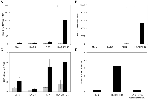
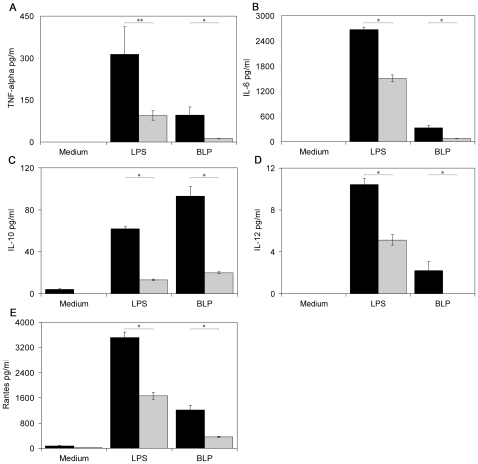
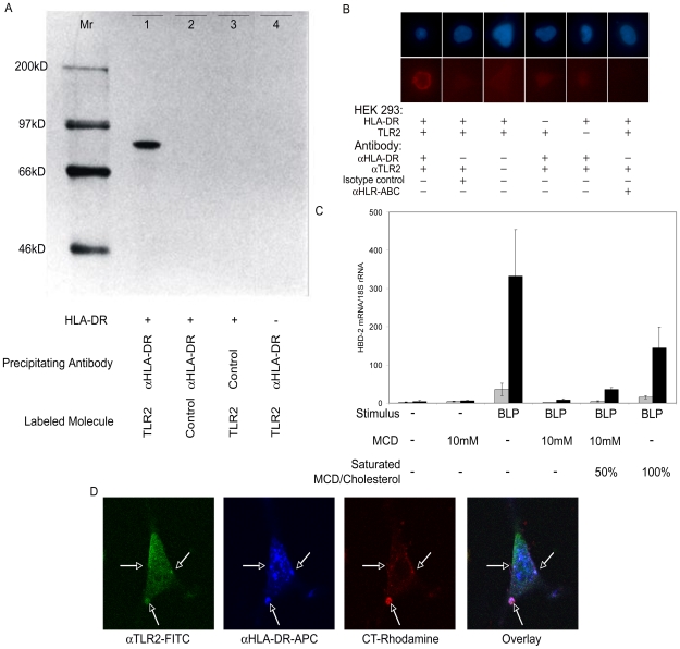
Methodology/principal findings: We found that co-expression of MHC class II molecules and TLR2 or TLR4 in human embryonic kidney (HEK) cells 293 leads to enhanced production of the anti-microbial peptide human-beta-defensin (hBD) 2 after treatment with TLR2 stimulus bacterial lipoprotein (BLP) or TLR4 ligand lipopolysaccharide (LPS), respectively. Furthermore, we found that peritoneal macrophages of MHC class II knock-out mice show a decreased responsiveness to TLR2 and TLR4 stimuli compared to macrophages of wild-type mice. Finally, we show that MHC class II molecules are physically and functionally associated with TLR2 in lipid raft domains of the cell membrane.
Conclusions/significance: These results demonstrate that MHC class II molecules are, in addition to their central role in adaptive immunity, also implicated in generating optimal innate immune responses.
Conflict of interest statement
Figures
References
-
- Takeda K, Kaisho T, Akira S. Toll-like receptors. Annu Rev Immunol. 2003;21:335–376. - PubMed
-
- Medzhitov R. Toll-like receptors and innate immunity. Nat Rev Immunol. 2001;1:135–145. - PubMed
-
- Akira S, Takeda K. Toll-like receptor signalling. Nature Reviews Immunology. 2004;4:499–511. - PubMed
-
- Docke WD, Randow F, Syrbe U, Krausch D, Asadullah K, et al. Monocyte deactivation in septic patients: restoration by IFN-gamma treatment. Nat Med. 1997;3:678–681. - PubMed
-
- Birchler T, Seibl R, Buchner K, Loeliger S, Seger R, et al. Human Toll-like receptor 2 mediates induction of the antimicrobial peptide human beta-defensin 2 in response to bacterial lipoprotein. Eur J Immunol. 2001;31:3131–3137. - PubMed
Publication types
MeSH terms
Substances
LinkOut - more resources
Full Text Sources
Other Literature Sources
Research Materials
Miscellaneous
