. 2010 Jan 20;5(1):e8791.
doi: 10.1371/journal.pone.0008791.

Endogenous morphine levels are increased in sepsis: a partial implication of neutrophils

Affiliations

Endogenous morphine levels are increased in sepsis: a partial implication of neutrophils

Elise Glattard et al. PLoS One. .

Abstract

Background: Mammalian cells synthesize morphine and the respective biosynthetic pathway has been elucidated. Human neutrophils release this alkaloid into the media after exposure to morphine precursors. However, the exact role of endogenous morphine in inflammatory processes remains unclear. We postulate that morphine is released during infection and can be determined in the serum of patients with severe infection such as sepsis.

Methodology: The presence and subcellular immunolocalization of endogenous morphine was investigated by ELISA, mass spectrometry analysis and laser confocal microscopy. Neutrophils were activated with Interleukin-8 (IL-8) or lipopolysaccharide (LPS). Morphine secretion was determined by a morphine-specific ELISA. mu opioid receptor expression was assessed with flow cytometry. Serum morphine concentrations of septic patients were determined with a morphine-specific ELISA and morphine identity was confirmed in human neutrophils and serum of septic patients by mass spectrometry analysis. The effects of the concentration of morphine found in serum of septic patients on LPS-induced release of IL-8 by human neutrophils were tested.

Principal findings: We confirmed the presence of morphine in human neutrophil extracts and showed its colocalisation with lactoferrin within the secondary granules of neutrophils. Morphine secretion was quantified in the supernatant of activated human polymorphonuclear neutrophils in the presence and absence of Ca(2+). LPS and IL-8 were able to induce a significant release of morphine only in presence of Ca(2+). LPS treatment increased mu opioid receptor expression on neutrophils. Low concentration of morphine (8 nM) significantly inhibited the release of IL-8 from neutrophils when coincubated with LPS. This effect was reversed by naloxone. Patients with sepsis, severe sepsis and septic shock had significant higher circulating morphine levels compared to patients with systemic inflammatory response syndrome and healthy controls. Mass spectrometry analysis showed that endogenous morphine from serum of patient with sepsis was identical to poppy-derived morphine.

Conclusions: Our results indicate that morphine concentrations are increased significantly in the serum of patients with systemic infection and that morphine is, at least in part, secreted from neutrophils during sepsis. Morphine concentrations equivalent to those found in the serum of septic patients significantly inhibited LPS-induced IL-8 secretion in neutrophils.


Conflict of interest statement

Competing Interests: The authors have declared that no competing interests exist.

Figures

Figure 1
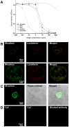
Figure 1. Evidence of the presence of morphine-like immunoreactivity in human neutrophils.
A . ELISA showing the specificity of the 6D6 antibody. B . Upper panel, double immunofluorescence confocal micrographs performed on purified neutrophils from healthy donors. Labeling was performed with a mouse anti-morphine antibody (visualized in green pseudocolor with a Cy5-conjugated IgG) and with an antibody against lactoferrin (a secondary granule marker) visualized in red pseudocolor with a Cy3-conjugated IgG. Colocalized immunolabelling (merged window) appears as yellow staining. Lower panel, higher magnification. C. Immunofluorescence confocal micrographs performed on a blood drop from an healthy donor. Labeling was performed with a mouse anti-morphine antibody (visualized in green pseudocolor with a Cy5-conjugated IgG). Phase contrast allows to observe both neutrophils and erythrocytes. Merged window correspond to the supperposition of the two pictures. D. To assess the specificity of the secondary antibody, control experiments were performed using Cy3-conjugated IgG or Cy5-conjugated IgG without primary antibody. Controls for morphine immunoreactivity were carried out by preincubating the antibody with morphine.
Figure 2
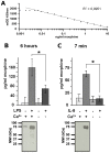
Figure 2. Morphine secretion from human primary neutrophils.
A . Typical dose-response curve of the morphine detection performed with the morphine-specific ELISA. B. Upper panel, Morphine secreted from neutrophils expressed as pg/ml. Morphine was quantified in culture medium after stimulating 20.106 neutrophils with LPS (10 ng/ml, 6 h; n = 6) in presence or absence of Ca2+. Basal secretion levels were obtained from neutrophils incubated without LPS (6 h; n = 6). Lower panel, Efficiency of secretion was assessed by monitoring the secretion of lactoferrin (75 kDa, a marker of secondary granules) by Western blot analysis. C. Upper panel , Morphine was quantified in culture medium after stimulating 20.106 neutrophils with IL-8 (50 nM, 7 min; n = 7) in presence or absence of Ca2+. Basal secretion levels were obtained from neutrophils incubated without IL-8 (7 min; n = 6). Lower panel, The efficiency of secretion was assessed by monitoring the secretion of lactoferrin (75 kDa, a marker of secondary granules) by Western blot analysis (50 µg). Amounts of secreted morphine in the LPS and IL-8 groups were statistically different from the two control groups (no LPS and IL-8; *: p<0.01 with a Mann-Whitney test after Bonferroni correction.
Figure 3
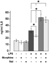
Figure 3. IL-8 secretion from human primary neutrophils.
IL-8 secreted from neutrophils expressed as ng/ml of morphine. Morphine was quantified in culture medium after stimulating 1.106 neutrophils with or without LPS (10 ng/ml, 24 h; n = 6), morphine 8 nM (2.5 ng/ml) and naloxone (10 µM). Basal secretion levels were obtained from neutrophils incubated without LPS (n = 6). *: p<0.05 with a Mann-Whitney test.
Figure 4
Figure 4. Detection of μ opioid receptor positive cells by and Western Blot, PCR, and flow cytometry.
A. Western blot analysis of 50 µg of SH-SY5Y (positive control) and human neutrophil extracts with the anti-μ opioid receptor (N-20). Control experiment shows the absence of cross reactivity for the secondary antibody. B. PCR amplification of MOR1 RNA performed on both naïve and LPS treated neutrophil total RNA extracts indicates the presence of a single band at 85 bp. Total RNA from SH-SY5Y cells is used as positive control, whereas negative control were done in absence of RNA and neutrophil RNA extract treated with DNAse. C. Flow cytometry with live gating on neutrophils using either a C-terminal antibody (C-20) or a N-terminal antibody (N-20). M1, M2 and M3 denote the different histogram gates used for determination of the median of fluorescence intensity D. Flow cytometry. Whole blood was stimulated with LPS (100 ng/ml) for different time intervals followed by labeling of μ opioid receptor expression on neutrophils with FITC-anti-μ opioid receptor (N-20). The percentage of cells showing an increased expression of μ opioid receptors is shown.
Figure 5
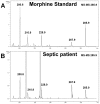
Figure 5. Characterization of morphine in serum of patients with sepsis.
Top, Q-TOF MS-MS analysis of morphine standard (286.17 Da; 1 pmol). Bottom, Q-TOF MS analysis of 1/50 of the morphine present in 10 ml of serum from a patient with sepsis.
Figure 6
Figure 6. Three-day follow up of morphine concentrations in infected and uninfected patients.
Boxplots of endogenous morphine concentrations measured in the serum of patients with sepsis (n = 13), severe sepsis (n = 8) and septic shock (n = 10), compared to critically ill patients with SIRS (n = 6) over 3 days of monitoring. Patients with sepsis, severe sepsis and septic shock had significantly higher serum concentrations of morphine than patients with SIRS (Sepsis vs. SIRS, p<0.05; severe sepsis vs SIRS, <0.01; septic shock vs SIRS, p<0.01).

Similar articles

Cited by

References

    1. Stefano GB, Goumon Y, Casares F, Cadet P, Fricchione GL, et al. Endogenous morphine. Trends Neurosci. 2000;23:436–442. - PubMed
    1. Gintzler AR, Gershon MD, Spector S. A nonpeptide morphine-like compound: immunocytochemical localization in the mouse brain. Science. 1978;199:447–448. - PubMed
    1. Goldstein A, Barrett RW, James IF, Lowney LI, Weitz CJ, et al. Morphine and other opiates from beef brain and adrenal. Proc Natl Acad Sci U S A. 1985;82:5203–5207. - PMC - PubMed
    1. Goumon Y, Laux A, Muller A, Aunis D. Central and Peripheral Endogenous Morphine. An R Acad Nac Farm. 2009;75:389–418.
    1. Stefano GB, Kream RM. Endogenous morphine synthetic pathway preceded and gave rise to catecholamine synthesis in evolution (Review). Int J Mol Med. 2007;20:837–841. - PubMed

Publication types