

doi: 10.1002/adma.200803786.
Highly Sensitive, Mechanically Stable Nanopore Sensors for DNA Analysis
Affiliations
- PMID: 20098720
- PMCID: PMC2808638
- DOI: 10.1002/adma.200803786
Item in Clipboard
Highly Sensitive, Mechanically Stable Nanopore Sensors for DNA Analysis
Adv Mater.
.
No abstract available
Figures
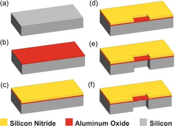
Process flow for the formation of Al2O3 nanopores. a) Start with double-side polished 300 μm thick Silicon wafer. b) Deposit 70 nm of Al2O3 by ALD. c) Deposit 500 nm low-stress SiN using PECVD process. d) Pattern 30 μm × 30 μm windows on the wafer front side via optical lithography and RIE. e) Pattern 30 μm × 30 μm windows on the wafer backside and etch using DRIE (SF6 + O2), and stop on the Al2O3 layer creating a membrane. f) Use a tightly focused electron beam to form nanometer-sized pores.
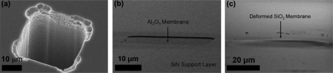
a) SEM image of backside trench formed using DRIE. b) SEM cross-section at 5° tilt of Al2O3 membrane with supporting SiN layer. Membrane is under low tensile stress and appears flat. c) SEM cross-section at 5° tilt of SiO2 membrane post-oxidation showing ∼5 μm vertical deflection over 50 μm span due to high compressive stress. Membrane is significantly deformed resulting in frequent failure.
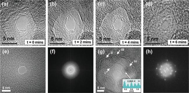
a–d) TEM phase-contrast images illustrating temporal contraction of an Al2O3 nanopore from an initial pore size of ∼4 nm to a final pore size of ∼1 nm. e) TEM phase-contrast image of 4 nm SiN pore. f) Corresponding FFT showing amorphous structure of SiN pore. g) TEM phase-contrast image of a 4 nm Al2O3 pore. Hexagonal Al2O3 nanocrystallites are shown by i, ii, iii, iv. iv is partially aligned with the zone axis with 2.28 Å atomic spacing corresponding to γ-Al2O3 in its 〈222〉 crystal orientation. h) Corresponding FFT showing polycrystalline structure of the pore.
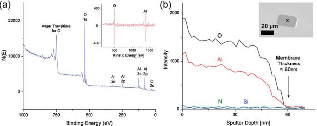
a) XPS results on Al2O3 films. Inset: Auger differential spectra of membrane region postrelease illustrating presence of only Al and O. b) Depth profiling using AES to extract membrane thickness of 60 ± 2 nm. Inset: tilted SEM image of membrane with marked region indicating Auger electron collection region.
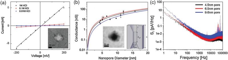
a) I–V characteristics of an 11.1 nm diameter pore measured in 10 mm KCl, 100 mm KCl, and 1 m KCl. Linear I–V characteristics suggest symmetric pore geometry. b) Pore conductance of 11 nanopores ranging in diameter from 4 to 16 nm. Red and blue lines represent conductance models from literature. Black line is a least-squares fit to the measured data. Predicted geometry of pore from conductance measurements. Left inset: thickness mapping of an 11.1 nm pore constructed using EFTEM. c) Power spectra of three different Al2O3 nanopores in 1 m KCl at 120 mV. Spectral components at high frequencies (f> 1 kHz) are significantly attenuated relative to Si3N4 and SiO2 systems.[14,15]
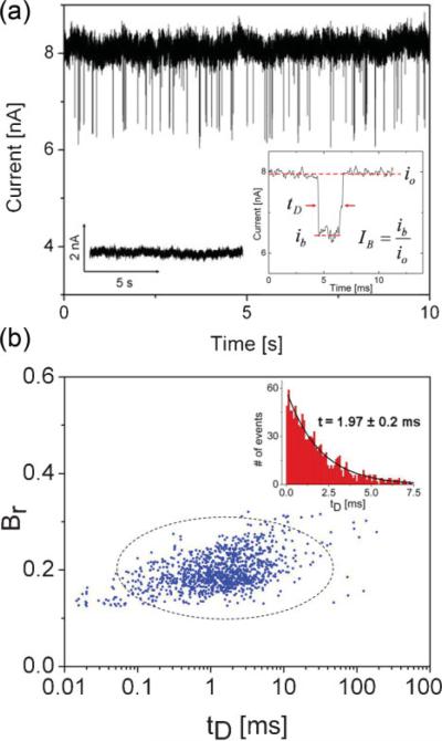
a) Typical current blockades seen in a 5.3 nm Al2O3 pore after the addition of 5 kbp dsDNA at a concentration of 6 nm at 500 mV. Data is low-pass filtered at 100 kHz. Left inset: negative control: pore current prior to the introduction of DNA is steady, no blockades are seen. Right inset: typical current blockade with annotations. b) Blockage ratio (Br) versus event dwell time (tD) for n = 1178 events. Primarily, a single blockade level with (Br) = 0.17 is seen. Inset: corresponding event-dwell-time histogram with time constant t = 1.97 ± 0.2 ms. Broad dwell-time distribution with large time constant suggests that events are DNA translocations rather than rapid collisions.
References
-
- Li J, Stein D, McMullan C, Branton D, Aziz MJ, Golovchenko JA. Nature. 2001;412:166. - PubMed
-
- Li J, Gershow M, Stein D, Brandin E, Golovchenko JA. Nat. Mater. 2003;2:611. - PubMed
-
- Storm AJ, Chen JH, Ling XS, Zandbergen HW, Dekker C. Nat. Mater. 2003;2:537. - PubMed
-
- Chang H, Kosari F, Andreadakis G, Alam MA, Vasmatzis G, Bashir R. Nano Lett. 2004;4:1551.
Grants and funding
LinkOut - more resources
Full Text Sources
Other Literature Sources
