. 2010 Jan 19;5(1):e8765.
doi: 10.1371/journal.pone.0008765.

Multiorgan detection and characterization of protease-resistant prion protein in a case of variant CJD examined in the United States

Affiliations

Multiorgan detection and characterization of protease-resistant prion protein in a case of variant CJD examined in the United States

Silvio Notari et al. PLoS One. .

Abstract

Background: Variant Creutzfeldt-Jakob disease (vCJD) is a prion disease thought to be acquired by the consumption of prion-contaminated beef products. To date, over 200 cases have been identified around the world, but mainly in the United Kingdom. Three cases have been identified in the United States; however, these subjects were likely exposed to prion infection elsewhere. Here we report on the first of these subjects.

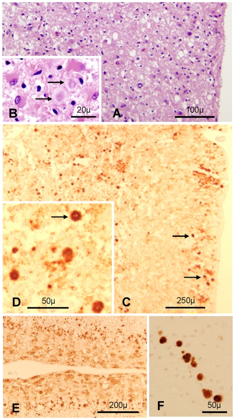
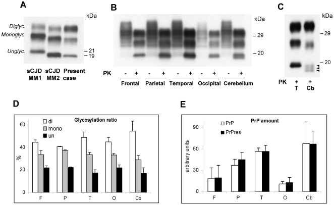
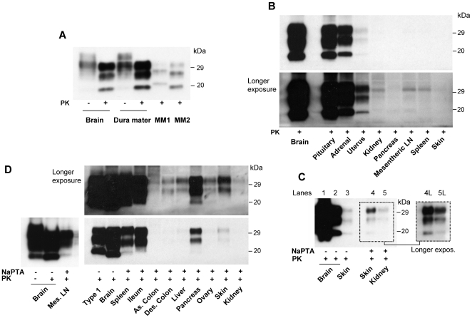
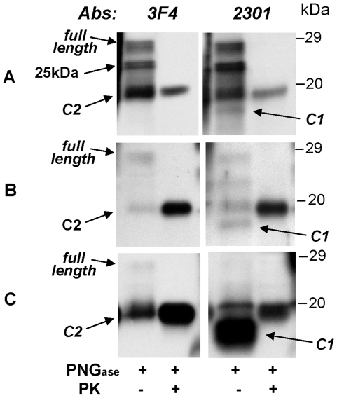
Methodology/principal findings: Neuropathological and genetic examinations were carried out using standard procedures. We assessed the presence and characteristics of protease-resistant prion protein (PrP(res)) in brain and 23 other organs and tissues using immunoblots performed directly on total homogenate or following sodium phosphotungstate precipitation to increase PrP(res) detectability. The brain showed a lack of typical spongiform degeneration and had large plaques, likely stemming from the extensive neuronal loss caused by the long duration (32 months) of the disease. The PrP(res) found in the brain had the typical characteristics of the PrP(res) present in vCJD. In addition to the brain and other organs known to be prion positive in vCJD, such as the lymphoreticular system, pituitary and adrenal glands, and gastrointestinal tract, PrP(res) was also detected for the first time in the dura mater, liver, pancreas, kidney, ovary, uterus, and skin.

Conclusions/significance: Our results indicate that the number of organs affected in vCJD is greater than previously realized and further underscore the risk of iatrogenic transmission in vCJD.


Conflict of interest statement

Competing Interests: The authors have declared that no competing interests exist.

Figures

Figure 1
Figure 1. Histological and immunohistochemical features.
A and B. The cerebral cortex is markedly disrupted with prominent astrogliosis and presence of amyloid plaques (arrows in B); frontal cortex; H.E. C and D: PrP immunostaining shows the presence of many plaques (arrows) and plaque-like aggregates in superficial and deep cortical regions with a punctate background staining; frontal cortex. E: Distinctive spot-like PrP immunostaining in the molecular layer and many plaques especially evident in the Purkinje cell layer of the cerebellum. F: Plaques, often in a row are present in the superficial white matter; cerebellum. C–F: Monoclonal antibody (mAb) 3F4.
Figure 2
Figure 2. Detection and characterization of PrPres and PK-sensitive PrP in brain.
A: Immunoblot of total homogenates (TH), treated with proteinase K (PK), obtained from the frontal cortex of sCJDMM1, sCJDMM2 (representing PrPres types 1 and 2, respectively) and the present case showing the over-representation of the upper band (Diglyc.) containing the diglycosylated form, and the co-migration of the lowest band (Unglyc.), containing the unglycosylated form, with the corresponding band of sCJDMM2. B: Immunoblot of TH from the four regions of the cerebral cortex and the cerebellum, treated with PK as indicated. The cerebellar unglycosylated PrPres isoform generates a thicker and overall slightly faster migrating band than the corresponding PrPres from the cerebral cortex. C: A high-resolution immunoblot (15%, 15 cm long gel) confirms that the monoglycosylated and unglycosylated PrPres isoforms from the cerebellum have a faster electrophoretic mobility than the corresponding forms from the cerebral cortex, and shows that the cerebellar unglycosylated isoform resolves into three fragments including a 19 kDa band, corresponding to PrPres type 2, and two additional bands of slightly lower relative molecular weight (arrowheads); T: Temporal; Cb: Cerebellum. In A–C membranes were probed with the mAb 3F4. D and E: Ratios of the PrPres glycoforms (D) and of the total PrP and PrPres (E) obtained from the same brain regions examined in panel B. Each bar represents the mean ± SD of three densitometric determinations on each of two tissue samples.
Figure 3
Figure 3. Detection of PrPres in non-nervous tissues.
A: PrPres from dura mater and frontal cortex (1∶24 dilution) from the present case is compared to PrPres of the frontal cortex from sCJDMM1 (type 1) and sCJDMM2 (type 2). B: Two film exposures of immunoblots from non-nervous tissues compared with that of the frontal cortex (1∶10 dilution). Pituitary gland, adrenal gland and uterus are clearly positive while the bands in the remaining preparations are considered to be non specific. C: PrPres from skin (double TH loading, equivalent to 4 mg of wet tissue) is barely detectable in TH (Lane 3) compared with frontal cortex TH, which is diluted 1∶4 (lane 1) or 1∶130 (lane 2). Skin PrPres is better detectable along with the kidney PrPres after sodium phosphotungstate (NaPTA) precipitation of PrPres (Lanes 4 and 5), especially after long exposure (Lanes 4L and 5L) (kidney TH loaded in double amount; probed with mAb 3F4). D: PrPres from mesenteric lymph nodes and other visceral organs recovered following NaPTA precipitation and compared with TH from the frontal cortex (1∶120 dilution) and sCJDMM1 following two film exposures. All organs but ascending colon are positive. Of note, unglycosylated isoform is underrepresented in all NaPTA precipitated samples as compared with that of the TH preparations (see panel A, lanes 3 and 4 and panel B). A–D: Membranes were probed with the mAb 3F4.
Figure 4
Figure 4. Characteristics of PK-sensitive PrP.
Immunoblot analysis of total homogenate from brain, pituitary gland and uterus are shown. The samples, with or without previous PK treatment, were deglycosylated with PNGase F. Membranes were probed with the mAbs 3F4 and 2301 as indicated. A: The brain has relatively large amounts of full-length isoform and PrPres C2 fragment but the N-terminus truncated PrPC fragment (C1) is poorly represented. In addition, brain preparation shows a previously unreported PK-sensitive fragment with molecular weight of 25 kDa (arrow), detectable only in deglycosylated samples, of undetermined origin. B: The C1 fragment is relatively better represented in the pituitary gland C: while it is overly abundant in the uterus.

Similar articles

Cited by

References

    1. Will RG, Ironside JW, Zeidler M, Cousens S, Estebeiro K, et al. A new variant of Creutzfeldt-Jakob disease in the UK. Lancet. 1996;347:921–925. - PubMed
    1. Will RG, Zeidler M, Stewart GE, Macleod MA, Ironside JW, et al. Diagnosis of new variant Creutzfeldt-Jakob disease. Ann Neurol. 2000;47:575–582. - PubMed
    1. Will RG, Ward HJ. Clinical features of variant Creutzfeldt-Jakob disease. Curr Top Microbiol Immunol. 2004;284:121–132. - PubMed
    1. Spencer MD, Knight RS, Will RG. First hundred cases of variant Creutzfeldt-Jakob disease: retrospective case note review of early psychiatric and neurological features. BMJ. 2002;324:1479–1482. - PMC - PubMed
    1. Collinge J, Sidle KCL, Meads J, Ironside J, Hill AF. Molecular analysis of prion strain variation and the aetiology of ‘new variant’ CJD. Nature. 1996;383:685–690. - PubMed

Publication types