. 2010 Jan 19;5(1):e8762.
doi: 10.1371/journal.pone.0008762.

Progression of Parkinson's disease pathology is reproduced by intragastric administration of rotenone in mice

Affiliations

Progression of Parkinson's disease pathology is reproduced by intragastric administration of rotenone in mice

Francisco Pan-Montojo et al. PLoS One. .

Abstract

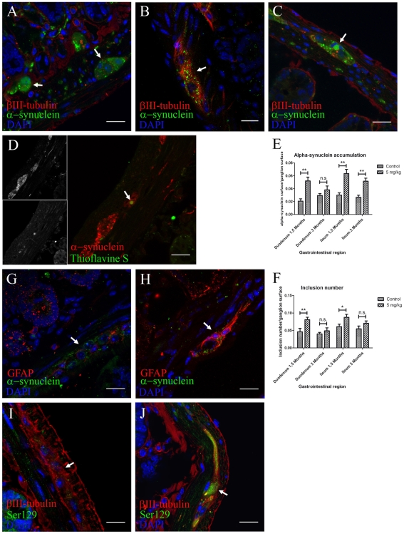
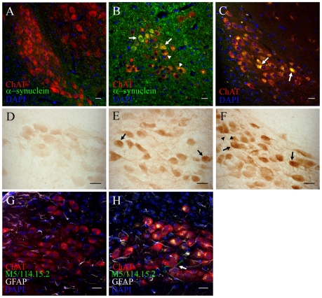
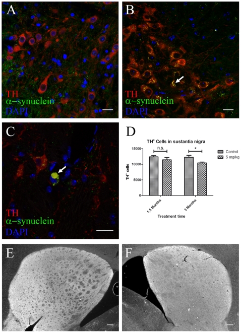
In patients with Parkinson's disease (PD), the associated pathology follows a characteristic pattern involving inter alia the enteric nervous system (ENS), the dorsal motor nucleus of the vagus (DMV), the intermediolateral nucleus of the spinal cord and the substantia nigra, providing the basis for the neuropathological staging of the disease. Here we report that intragastrically administered rotenone, a commonly used pesticide that inhibits Complex I of the mitochondrial respiratory chain, is able to reproduce PD pathological staging as found in patients. Our results show that low doses of chronically and intragastrically administered rotenone induce alpha-synuclein accumulation in all the above-mentioned nervous system structures of wild-type mice. Moreover, we also observed inflammation and alpha-synuclein phosphorylation in the ENS and DMV. HPLC analysis showed no rotenone levels in the systemic blood or the central nervous system (detection limit [rotenone]<20 nM) and mitochondrial Complex I measurements showed no systemic Complex I inhibition after 1.5 months of treatment. These alterations are sequential, appearing only in synaptically connected nervous structures, treatment time-dependent and accompanied by inflammatory signs and motor dysfunctions. These results strongly suggest that the local effect of pesticides on the ENS might be sufficient to induce PD-like progression and to reproduce the neuroanatomical and neurochemical features of PD staging. It provides new insight into how environmental factors could trigger PD and suggests a transsynaptic mechanism by which PD might spread throughout the central nervous system.


Conflict of interest statement

Competing Interests: Francisco Pan-Montojo has a patent application pending for this animal model (Application number PCT/EP 2009/005688). This does not alter the authors' adherence to all the PLoS ONE policies on sharing data and materials, as detailed online in the guide for authors (http://www.plosone.org/static/policies.action#sharing).

Figures

Figure 1
Figure 1. Locally administered rotenone induces alpha-synuclein phosphorylation, accumulation and aggregation with gliosis in ENS ganglia.
(scale bars 20 µm). A, B, C, anti βIII-tubulin, alpha-synuclein and DAPI staining in duodenum (B) and ileum (A,C) sections. Arrow in B, 1.5 months treatment induced an increased alpha-synuclein punctate pattern inside enteric nervous system ganglia when compared to 3 months controls (A). Arrow in C, 3 months treatment induced formation of larger alpha-synuclein inclusions (|>6 µm). D, immunofluorescence staining using anti-alpha-synuclein, Thioflavine S and DAPI. Arrow in D, only 3 month treated mice showed aggregation of these larger alpha-synuclein accumulations. E, F, quantification of the experiment shown in AC was made using automatic segmentation and entropy-based thresholding methods. Single-asterisk, P<0.05, and double-asterisk, P<0.01. E, each column represents total alpha-synuclein surface/ganglion surface. F, each column represents total number of alpha-synuclein inclusions/ganglion surface. All graphs show mean ± s.e.m. G, H, max-projection of staining against GFAP, alpha-synuclein and DAPI on duodenum sections from control (G) and treated (H) mice. I, J, max-projection of anti-βIII-tubulin, anti-phospho-alpha-synuclein (Ser 129) and DAPI staining on duodenum sections from control (I) and treated (J) animals.
Figure 2
Figure 2. Intracellular and axonal alpha-synuclein increases in the intermediolateral nucleus and the dorsal horn lamina I layer of the spinal cord after oral rotenone treatment.
(scale bars 20 µm) A, B, C, Immunostaining against alpha-synuclein and choline acetyl transferase (ChAT) in spinal cord sections showing the intermediolateral nucleus ChAT+ neurons from 3 months control mice (A), 1.5 months (B) and 3 months (C) treated mice. Arrow in B, colocalization of increased intracellular alpha-synuclein and ChAT+ stainings in the IML. Arrow in C, large alpha-synuclein inclusion (|>7.5 µm) inside an IML ChAT+ neuron. DE, fluorescence intensity color-coded images from 3 months control (D, D') and 3 months treated mice (E, E') spinal cord sections stained using DAPI and alpha-synuclein and ChAT antibodies. Arrows in D and E, areas in the proximity of ChAT+ neurons. F, mean fluorescence quantification of experiment shown in D and E. Double asterisk, P<0,01. Columns represent mean alpha-synuclein fluorescence in and around ChAT+ neurons in the IML/mean alpha-synuclein fluorescence in the region anterior to the IML. Graph shows mean ± s.e.m.. G, H, DAB-staining against apha-synuclein using synuclein-1 antibody in the dorsal horn of the spinal cord from 3 months treated (H) and control (G) mice. Arrows in GH, lamina I layer of the dorsal horn.
Figure 3
Figure 3. Intragastrically administered rotenone induces alpha-synuclein accumulation, oxidative stress and inflammation in the dorsal motor nucleus vagus.
(scale bars 20 µm). A, B, double-immunofluorescence staining against alpha-synuclein and ChAT on DMV sections from 1.5 months control (A) and 1.5 months treated (B) mice. Arrows in B, increased intracellular alpha-synuclein in DMV neurons already after 1.5 months. Arrowheads in B, autofluorescent punctate inclusion pattern inside ChAT+ neurons. C, DMV sections stained with ChAT and DAPI were sequentially excited with 488 and 561 laser wavelengths. Arrows in C, large intracellular auto-fluorescent inclusions inside ChAT+ neurons of the DMV (arrows). D, E, F, Light microscopy images of alpha-synuclein staining from 1.5 months control (D), 1.5 months (E) and 3 months (F) treated mice. Arrows in E and F, increased staining intensity inside DMV neuronal soma in treated mice. Arrowheads in F, increased alpha-synuclein staining inside neuronal processes G, H, average-projection of triple-immunofluorescence staining against ChAT, GFAP, MHC II (clone M5/114.15.2) and DAPI on sections from control (G) and treated (H) mice after 3 month treatment. Arrow in H, activated microglial cell in the DMV.
Figure 4
Figure 4. Alpha-synuclein accumulation and neuronal loss in the SNc after 3 but not 1.5 months intragastrical rotenone treatment.
(AC, scale bars 20 µm; EF, scale bars 200 µm). A, B, C, immunostaining against TH, alpha-synuclein and DAPI on SNc sections from 1.5 months control (A) and 3 months (BC) treated mice. Arrow in B, alpha-synuclein small inclusions inside TH+ neurons. Arrow in C, large alpha-synuclein inclusion (|>8.14 µm) inside a dopamineric neuron in the SN. D, stereological quantification (n = 3) of TH+ neurons in the SN from control and treated mice. Asterisk, P<0.05. Number of neurons was determined based on the optical fractionator principle using StereoInvestigator software (MicroBrightField Inc., Williston, USA). Each column represents total number of TH+ neurons in the SN in 1.5 and 3 months control and treated mice. Graph shows mean ± s.e.m. E, F, TH immunostaining on striatum in 1.5 months control (E) and 3 months treated (F) mice.

Similar articles

Cited by

References

    1. de Rijk MC, Launer LJ, Berger K, Breteler MM, Dartigues JF, et al. Prevalence of Parkinson's disease in Europe: A collaborative study of population-based cohorts. Neurologic Diseases in the Elderly Research Group. Neurology. 2000;54:S21–23. - PubMed
    1. Litvan I, Bhatia KP, Burn DJ, Goetz CG, Lang AE, et al. Movement Disorders Society Scientific Issues Committee report: SIC Task Force appraisal of clinical diagnostic criteria for Parkinsonian disorders. Mov Disord. 2003;18:467–486. - PubMed
    1. Crossman AR. Neural mechanisms in disorders of movement. Comp Biochem Physiol A Comp Physiol. 1989;93:141–149. - PubMed
    1. Albin RL, Young AB, Penney JB. The functional anatomy of basal ganglia disorders. Trends Neurosci. 1989;12:366–375. - PubMed
    1. Lang AE, Obeso JA. Time to move beyond nigrostriatal dopamine deficiency in Parkinson's disease. Ann Neurol. 2004;55:761–765. - PubMed

Publication types