. 2010 Jan 19;5(1):e8769.
doi: 10.1371/journal.pone.0008769.

Mycobacterial nucleoside diphosphate kinase blocks phagosome maturation in murine RAW 264.7 macrophages

Affiliations

Mycobacterial nucleoside diphosphate kinase blocks phagosome maturation in murine RAW 264.7 macrophages

Jim Sun et al. PLoS One. .

Abstract

Background: Microorganisms capable of surviving within macrophages are rare, but represent very successful pathogens. One of them is Mycobacterium tuberculosis (Mtb) whose resistance to early mechanisms of macrophage killing and failure of its phagosomes to fuse with lysosomes causes tuberculosis (TB) disease in humans. Thus, defining the mechanisms of phagosome maturation arrest and identifying mycobacterial factors responsible for it are key to rational design of novel drugs for the treatment of TB. Previous studies have shown that Mtb and the related vaccine strain, M. bovis bacille Calmette-Guérin (BCG), disrupt the normal function of host Rab5 and Rab7, two small GTPases that are instrumental in the control of phagosome fusion with early endosomes and late endosomes/lysosomes respectively.

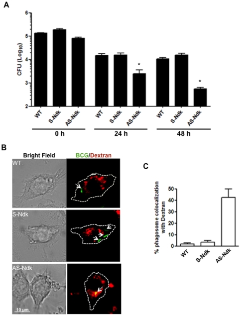
Methodology/principal findings: Here we show that recombinant Mtb nucleoside diphosphate kinase (Ndk) exhibits GTPase activating protein (GAP) activity towards Rab5 and Rab7. Then, using a model of latex bead phagosomes, we demonstrated that Ndk inhibits phagosome maturation and fusion with lysosomes in murine RAW 264.7 macrophages. Maturation arrest of phagosomes containing Ndk-beads was associated with the inactivation of both Rab5 and Rab7 as evidenced by the lack of recruitment of their respective effectors EEA1 (early endosome antigen 1) and RILP (Rab7-interacting lysosomal protein). Consistent with these findings, macrophage infection with an Ndk knocked-down BCG strain resulted in increased fusion of its phagosome with lysosomes along with decreased survival of the mutant.

Conclusion: Our findings provide evidence in support of the hypothesis that mycobacterial Ndk is a putative virulence factor that inhibits phagosome maturation and promotes survival of mycobacteria within the macrophage.


Conflict of interest statement

Competing Interests: The authors have declared that no competing interests exist.

Figures

Figure 1
Figure 1. Ndk interacts with and deactivates Rab5 and Rab7 GTPases. A and B.
ELISA microplates were coated with 10 µg/ml Mtb or M. smegmatis Ndk or control BSA, and incubated for 1 h with increasing concentrations of Rab5 or Rab7, previously loaded with 1 mM GTP in reaction buffer (50 mM HEPES pH 7.4, 50 mM NaCl, 0.1 mM DTT, 5 mM EDTA and 1 mg/ml BSA) for 10 min at 37°C. Following 3 washes, attached Rab5 or Rab7 was probed with primary rabbit anti-Rab5 or Rab7, followed by secondary anti-rabbit-HRP conjugate. Thereafter, the interaction was visualized at absorbance 450 nm after addition of TMB substrate. Values from control BSA were subtracted. Results (mean ± s.e.m) are from 3 independent experiments. C and D. Recombinant Ndk (3 µg) and GTP-loaded Rab5 or Rab7 (3 µg) were incubated in PBS buffer for 1 h at 4°C. Thereafter, anti-Ndk antibodies (1∶100) were added (1 h at 4°C) and subjected to immunoprecipitation with protein A agarose beads for 30 min at room temperature. Samples were washed three times then analyzed by SDS-PAGE and western blot with anti-Ndk and anti-Rab5 or anti-Rab7 followed subsequently by monoclonal anti-rabbit IgG, native-peroxidase. Pulled down Rab7 and Rab5 are shown as the 25 kDa and 27 kDa protein bands respectively, while the 15 kDa protein band correspond to Ndk. E and F. Recombinant Rab5 and Rab7 were loaded with [γ-32P]-GTP and spotted onto nitrocellulose membranes. After extensive washes, membranes were either left untreated or incubated with recombinant Mtb or M. smegmatis Ndk at room temperature for 2 h. Membranes were washed, dried and exposed to X-ray film (upper panel). The radioactive signal observed depicts remaining active GTP-Rab7 or -Rab5 on membranes and values are quantification of bound [γ-32P]-GTP relative to control untreated samples as determined by radioactive count in a liquid scintillation counter. After film development, membranes were probed with anti-Rab7 to ensure equal spotting of Rab7 protein (lower panel).
Figure 2
Figure 2. Ndk contributes to phagosome maturation arrest.
A. RAW 264.7 cells were pulse-chased overnight with FITC-DXT (0.5 mg/ml) and allowed to ingest control BSA-coated, Mtb Ndk-coated, or M. smegmatis Ndk-coated latex beads. Two hours post-phagocytosis, cells were washed and fixed for analysis by confocal microscopy. B. Quantification of the confocal data shown in panel A. C. RAW 264.7 cells were loaded with FITC-DXT overnight and allowed to ingest a mixture (1∶1) of either PKH-labelled BSA-beads and unlabelled BSA-beads (upper panel, control) or PKH-labelled BSA-beads and unlabelled Ndk-beads (lower panel). Two hours post-phagocytosis, cells were trypsinized, washed, and fixed for analysis by confocal microscopy. The top panel (BSA control), the yellow signal reflects a colocalization of the PKH-BSA-beads with the lysosomal marker dextran. In the bottom panel dotted circles indicate the position of Ndk-beads, while the red fluorescent signal (PKH) shows the location of BSA-beads and a significant decrease of dextran colocalization with distant PKH-BSA-beads. D. Quantification of the confocal data shown in panel C. Values in B and D are the mean ± SD of phagosome colocalization of with FITC-DXT in 50–80 cells from three independent experiments.
Figure 3
Figure 3. Ndk inhibits EEA1 recruitment to phagosomes.
A. RAW cells were transfected with EEA1-GFP and thereafter allowed to ingest either control BSA- or Ndk-coated latex beads. The green signal shows presence of EEA1 on the phagosome containing control beads (upper panel), while the lack of signal around the phagosome (lower panel) shows diminished recruitment of EEA1 to Ndk-bead containing phagosomes. B. Quantification of the confocal data shown in panel A. C. Raw cells were transfected with 2xFYVE-GFP, and thereafter allowed to phagocytose either control BSA- or Ndk-coated latex beads. 2xFYVE is a specific marker for PI3P. The green signal seen in the upper control panel indicates an abundance of PI3P generated on the phagosome, while the lack of signal on Ndk-bead phagosomes indicate absence of PI3P. D. Quantification of confocal data shown in panel C. Values in B and D are the mean ± SD of phagosome colocalization with EEA1-GFP and 2xFYVE-GFP respectively in 50–80 cells from three independent experiments.
Figure 4
Figure 4. A. Ndk disrupt Rab7-RILP interaction.
RAW cells were double transfected with Rab7-GFP and RILP-DsRed as described in Materials and Methods. Thereafter, cells were allowed to ingest either control BSA- or Ndk-coated latex beads. The yellow signal seen (upper panel) shows colocalization of Rab7 and RILP on the phagosome. The green signal (lower panel) shows phagosomes positive for Rab7 but no recruitment of RILP. B. Quantification of the confocal data shown in panel A. Values in B are the mean ± SD of phagosome colocalization with RILP-DsRed in 50–80 cells from three independent experiments.
Figure 5
Figure 5. Generation of recombinant BCG with knocked down Ndk expression.
A. Wild-type BCG, BCG transformed with pMV261-S-Ndk (sense, overexpression), and BCG transformed with pMV261-AS-Ndk (antisense, knockdown) were lysed as described in Materials and Methods, and subjected to 15% SDS-PAGE, followed by western blot with anti-Ndk antibodies. Mycobacterial lipoamide dehydrogenase C (LpdC) was used as an internal control for equal loading. B. Growth curve of the BCG strains shown in panel A expressed as Absorbance at 600 nm.
Figure 6
Figure 6. BCG-AS−Ndk has decreased intracellular survival due to increased phagolysosome fusion.
A. RAW macrophages were infected with BCG strains (MOI of 10∶1). Then culture media was supplemented with 50 µg/ml gentamicin to kill extracellular non-ingested mycobacteria. Cells were washed three times in PBS and lysed in 0.025% SDS 1 h (0 h time point), 24 h, and 48 h post-infection. Serial dilutions of recovered bacteria were then plated on solid 7H10 media supplemented with 10% OADC. CFU counts were performed after 3 weeks incubation at 37°C,. Bars, mean ± s.e.m. (three independent experiments). B. RAW cells adherent to cover slips were loaded with 0.5 mg/ml Texas Red-Dextran overnight and then infected with FITC-labelled BCG strains (wild-type: WT, expression of sense (S-), or antisense (AS-) Ndk) at an MOI of 20∶1. At 4 h post-phagocytosis, cells were fixed with 2.5% paraformaldehyde and mounted onto slides for confocal analysis. Bright field and merged fluorescent images are shown with an outline of the cell boundaries. Green signal indicates BCG that are not colocalized with dextran, while yellow signal shows colocalization of BCG with lysosomes. C. Quantification of data shown in panel B.
Figure 7
Figure 7. Sequence alignment of Mtb, BCG, and M. smegmatis.
There is 100% homology between Mtb and BCG Ndk, which decreases to ∼80% when compared to M. smegmatis Ndk. Residues of difference that could potentially be important in the catalytic GAP activity are highlighted.

Similar articles

Cited by

References

    1. North RJ, Jung YJ. Immunity to tuberculosis. Annu Rev Immunol. 2004;22:599–623. - PubMed
    1. LoBue P. Extensively drug-resistant tuberculosis. Curr Opin Infect Dis. 2009;22(2):167–173. - PubMed
    1. Cooper AM. Cell-mediated immune responses in tuberculosis. Annu Rev Immunol. 2009;27:393–422. - PMC - PubMed
    1. Jozefowski S, Sobota A, Kwiatkowska K. How mycobacterium tuberculosis subverts host immune responses. Bioessays. 2008;30(10):943–954. - PubMed
    1. Lee WL, Gold B, Darby C, Brot N, Jiang X, et al. Mycobacterium tuberculosis expresses methionine sulphoxide reductases A and B that protect from killing by nitrite and hypochlorite. Mol Microbiol. 2009;71(3):583–593. - PMC - PubMed

Publication types

Substances