. 2010 Jan 20;5(1):e8788.
doi: 10.1371/journal.pone.0008788.

Brainstem circuitry regulating phasic activation of trigeminal motoneurons during REM sleep

Affiliations

Brainstem circuitry regulating phasic activation of trigeminal motoneurons during REM sleep

Christelle Anaclet et al. PLoS One. .

Abstract

Background: Rapid eye movement sleep (REMS) is characterized by activation of the cortical and hippocampal electroencephalogram (EEG) and atonia of non-respiratory muscles with superimposed phasic activity or twitching, particularly of cranial muscles such as those of the eye, tongue, face and jaw. While phasic activity is a characteristic feature of REMS, the neural substrates driving this activity remain unresolved. Here we investigated the neural circuits underlying masseter (jaw) phasic activity during REMS. The trigeminal motor nucleus (Mo5), which controls masseter motor function, receives glutamatergic inputs mainly from the parvocellular reticular formation (PCRt), but also from the adjacent paramedian reticular area (PMnR). On the other hand, the Mo5 and PCRt do not receive direct input from the sublaterodorsal (SLD) nucleus, a brainstem region critical for REMS atonia of postural muscles. We hypothesized that the PCRt-PMnR, but not the SLD, regulates masseter phasic activity during REMS.

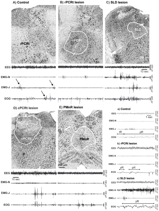
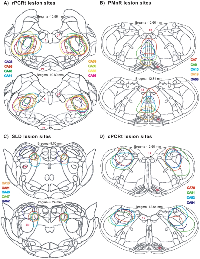
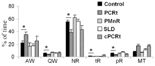
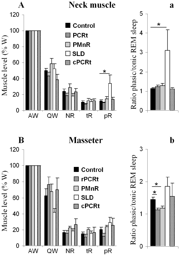
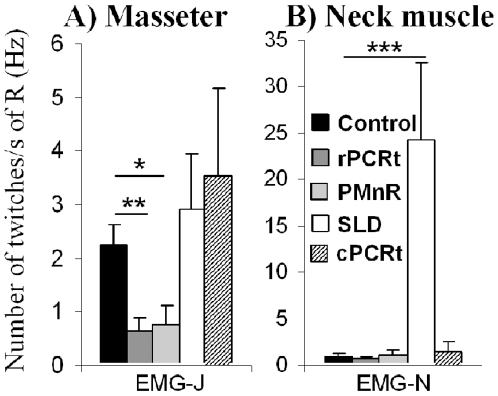
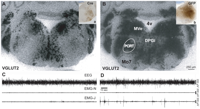
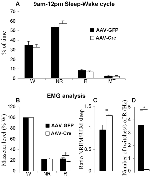
Methodology/principal findings: To test our hypothesis, we measured masseter electromyogram (EMG), neck muscle EMG, electrooculogram (EOG) and EEG in rats with cell-body specific lesions of the SLD, PMnR, and PCRt. Bilateral lesions of the PMnR and rostral PCRt (rPCRt), but not the caudal PCRt or SLD, reduced and eliminated REMS phasic activity of the masseter, respectively. Lesions of the PMnR and rPCRt did not, however, alter the neck EMG or EOG. To determine if rPCRt neurons use glutamate to control masseter phasic movements, we selectively blocked glutamate release by rPCRt neurons using a Cre-lox mouse system. Genetic disruption of glutamate neurotransmission by rPCRt neurons blocked masseter phasic activity during REMS.

Conclusions/significance: These results indicate that (1) premotor glutamatergic neurons in the medullary rPCRt and PMnR are involved in generating phasic activity in the masseter muscles, but not phasic eye movements, during REMS; and (2) separate brainstem neural circuits control postural and cranial muscle phasic activity during REMS.


Conflict of interest statement

Competing Interests: The authors have declared that no competing interests exist.

Figures

Figure 1
Figure 1. Lesion histology and REM sleep phenotype following cell-body specific lesions in rats.
(A) unlesioned control, the arrows show masseter twitches (EMG-J) and bursting in the EOG during REM sleep (B) rPCRt lesion (OX-SAP,0.1%, 330 nl; rPCRt is defined as the portion of PCRt that is rostral to the inferior olive and caudal to the facial tract; cPCRt extends caudally from this), (C) SLD lesion (OX-SAP,0.1%, 130 nl), (D) cPCRt lesion (OX-SAP,0.1%, 330 nl) and (E) PMnR lesion (OX-SAP,0.1%, 230 nl). For each experimental condition a 2 min REM sleep episode example (from the 256 Hz recording) is shown to illustrate the typical EEG, EMG-neck, EMG-jaw and EOG recordings. A set of extended recording examples (from the 256 Hz recording) is showed in the lower right panel and includes: a) control, b) rPCRt lesion and c) SLD lesion cases. Note that during REM sleep, (1) control rats showed activated EEG, EMG-N atonia, EMG-J phasic twitches and rapid eye movements; (2) rPCRt and PMnR lesions eliminated/reduced EMG-J twitches; (3) SLD lesions increased phasic activity of the neck muscles; and (4) cPCRt lesion did not alter the REM sleep EEG/EMGs/EOG. SLD: sublaterodorsal nucleus; PCRt: parvocellular reticular nucleus; PMnR: paramedian reticular area; PB: parabrachial nucleus; Ve: vestibular nucleus; 7: facial nucleus; IO: inferior olive; EEG: electroencephalogram; EMG-N: neck electromyogram; EMG-J: masseter or jaw electromyogram; EOG: electrooculogram; tR: tonic REM sleep; pR: phasic REM sleep.
Figure 2
Figure 2. Lesion maps for all rPCRt(A), PMnR(B), SLD(C) and cPCRt (D) cases in rats.
5N: trigeminal nucleus; 6: abducens nucleus; 12: hypoglossal nucleus; g7: facial nerve; IO: inferior olive; Py: pontine nuclei.
Figure 3
Figure 3. Three hours (9am-12pm) sleep-wake distribution following cell-body specific lesions in rats.
Percentage of time of AW: active wake, QW: quiet wake, NR: non REM sleep, tR: tonic REM sleep, pR: phasic REM sleep and MT: artifacts during the 3 h, 2 kHz recording for EMG analysis. All values are means ± SEM; *p<0.05 using a two-tailed t-test.
Figure 4
Figure 4. Neck muscle and masseter activity following cell-body specific lesions in rats.
In A (neck) and B (jaw), muscle activity for each stage is expressed in percentage of the active wake muscle activity (left panel). a and b (right panel) represent corresponding ratios of phasic and tonic REM sleep muscle activity. AW: active wake; QW: quiet wake; NR: non REM sleep; tR: tonic REM sleep; pR: phasic REM sleep. All values are means ± SEM; *p<0.05 using a two-tailed t-test.
Figure 5
Figure 5. Neck muscle and masseter twitches during REM sleep following lesions in rats.
Y axis = number of muscle twitches/REM sleep time (s). Note that SLD lesion significantly increase neck muscle twitches while both PMnR and rPCRt lesions significantly decrease masseter twitches, as compared with the control groups. EMG-J: jaw or masseter electromyogram; EMG-N: neck electromyogram; R: total REM sleep; All values are means ± SEM; *p<0.05; **p<0.01; ***p<0.001 using a two-tailed t-test.
Figure 6
Figure 6. Selective elimination of glutamate release from the rPCRt.
Injections (boxes in upper right corners) of AAV-Cre or AAV-GFP encompassing the rPCRt are shown by Cre (a, knockout) or GFP (b, control) immunostaining in a floxed Vglut2 mouse. VGLUT2 mRNA signal (black puncta) in an AAV-Cre injected mouse (A) and AAV-GFP injected mouse (B) in the region corresponding with Cre or GFP immunolabelling in the Vglut2 mouse. Note the loss of VGLUT2 mRNA in the Cre, but not GFP, injected animal. C & D, EEG, EMG-Neck and EMG-Jaw traces during REM sleep in an AAV-Cre injected VGLUT2 floxed mouse (C) and AAV-GFP (vector control) injected VGLUT2 floxed mouse (D). Specific elimination of Vglut2 from the rPCRt neurons resulted in reduced phasic masseter twitches during REM sleep.
Figure 7
Figure 7. Sleep-wake and masseter activity analysis following loss of rPCRt glutamate release.
(A) Percentage of W: wake, NR: non REM sleep, R: REM sleep and MT: artifacts from the 2 kHz recording for EMG (3 hours, 9AM-12PM). (B) Masseter activity of each stage expressed as a percentage of wake masseter activity in AAV-Cre and AAV-GFP (control) injected mice. (C) Ratio of NR and REM sleep masseter activity in AAV-Cre and AAV-GFP injected mice. (D) masseter twitches during REM sleep in AAV-Cre and AAV-GFP injected mice. Y axis = number of muscle twitches/REM sleep time (s). All values are means ± SEM; *p<0.05 using a two-tailed t-test.

Similar articles

Cited by

References

    1. Jouvet M, Michel F, Courjon J. [On a stage of rapid cerebral electrical activity in the course of physiological sleep.]. C R Seances Soc Biol Fil. 1959;153:1024–1028. - PubMed
    1. Jouvet M, Michel F. [Electromyographic correlations of sleep in the chronic decorticate & mesencephalic cat.]. C R Seances Soc Biol Fil. 1959;153:422–425. - PubMed
    1. Aserinsky E, Kleitman N. Regularly occurring periods of eye motility, and concomitant phenomena, during sleep. Science. 1953;118:273–274. - PubMed
    1. Kato T, Masuda Y, Kanayama H, Morimoto T. Muscle activities are differently modulated between masseter and neck muscle during sleep-wake cycles in guinea pigs. Neurosci Res. 2007;58:265–271. - PubMed
    1. Burgess C, Lai D, Siegel J, Peever J. An endogenous glutamatergic drive onto somatic motoneurons contributes to the stereotypical pattern of muscle tone across the sleep-wake cycle. J Neurosci. 2008;28:4649–4660. - PMC - PubMed

Publication types