

Methamphetamine self-administration is associated with persistent biochemical alterations in striatal and cortical dopaminergic terminals in the rat
- PMID: 20098750
- PMCID: PMC2808335
- DOI: 10.1371/journal.pone.0008790
Methamphetamine self-administration is associated with persistent biochemical alterations in striatal and cortical dopaminergic terminals in the rat
Abstract
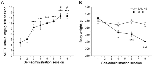
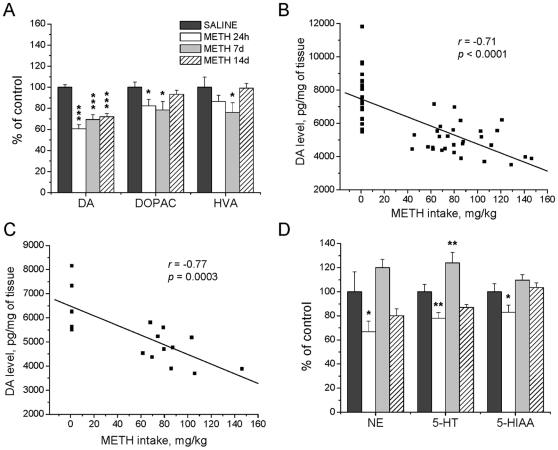
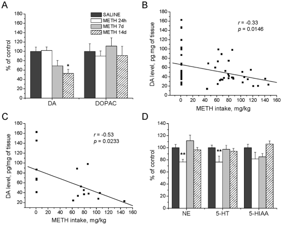
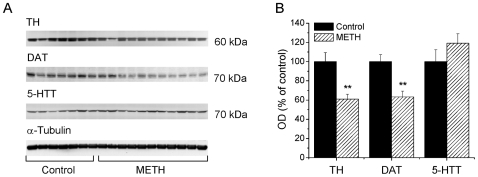
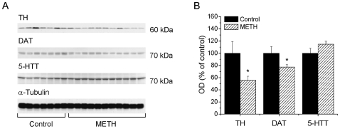
Methamphetamine (meth) is an illicit psychostimulant that is abused throughout the world. Repeated passive injections of the drug given in a single day or over a few days cause significant and long-term depletion of dopamine and serotonin in the mammalian brain. Because meth self-administration may better mimic some aspects of human drug-taking behaviors, we examined to what extent this pattern of drug treatment might also result in damage to monoaminergic systems in the brain. Rats were allowed to intravenously self-administer meth (yoked control rats received vehicle) 15 hours per day for 8 days before being euthanized at either 24 hours or at 7 and 14 days after cessation of drug taking. Meth self-administration by the rats was associated with a progressive escalation of daily drug intake to 14 mg/kg per day. Animals that self-administered meth exhibited dose-dependent decreases in striatal dopamine levels during the period of observation. In addition, there were significant reductions in the levels of striatal dopamine transporter and tyrosine hydroxylase proteins. There were also significant decreases in the levels of dopamine, dopamine transporter, and tyrosine hydroxylase in the cortex. In contrast, meth self-administration caused only transient decreases in norepinephrine and serotonin levels in the two brain regions, with these values returning to normal at seven days after cessation of drug taking. Importantly, meth self-administration was associated with significant dose-dependent increases in glial fibrillary acidic protein in both striatum and cortex, with these changes being of greater magnitude in the striatum. These results suggest that meth self-administration by rats is associated with long-term biochemical changes that are reminiscent of those observed in post-mortem brain tissues of chronic meth abusers.
Conflict of interest statement
Figures

Similar articles
-
Methamphetamine self-administration causes persistent striatal dopaminergic alterations and mitigates the deficits caused by a subsequent methamphetamine exposure.J Pharmacol Exp Ther. 2012 Feb;340(2):295-303. doi: 10.1124/jpet.111.188433. Epub 2011 Oct 27. J Pharmacol Exp Ther. 2012. PMID: 22034657 Free PMC article.
-
Epothilone D prevents binge methamphetamine-mediated loss of striatal dopaminergic markers.J Neurochem. 2016 Feb;136(3):510-25. doi: 10.1111/jnc.13391. Epub 2015 Dec 10. J Neurochem. 2016. PMID: 26465779 Free PMC article.
-
Methamphetamine-induced changes in the object recognition memory circuit.Neuropharmacology. 2012 Feb;62(2):1119-26. doi: 10.1016/j.neuropharm.2011.11.003. Epub 2011 Nov 18. Neuropharmacology. 2012. PMID: 22115899 Free PMC article.
-
Detection of methamphetamine neurotoxicity in forensic autopsy cases.Leg Med (Tokyo). 2009 Apr;11 Suppl 1:S63-5. doi: 10.1016/j.legalmed.2009.01.003. Epub 2009 Mar 6. Leg Med (Tokyo). 2009. PMID: 19269222 Review.
-
Methamphetamine addiction: involvement of CREB and neuroinflammatory signaling pathways.Psychopharmacology (Berl). 2016 May;233(10):1945-62. doi: 10.1007/s00213-016-4235-8. Epub 2016 Feb 12. Psychopharmacology (Berl). 2016. PMID: 26873080 Free PMC article. Review.
Cited by
-
Mitochondrial oxidant stress mediates methamphetamine neurotoxicity in substantia nigra dopaminergic neurons.Neurobiol Dis. 2021 Aug;156:105409. doi: 10.1016/j.nbd.2021.105409. Epub 2021 May 31. Neurobiol Dis. 2021. PMID: 34082123 Free PMC article.
-
Effects of drugs of abuse on hippocampal plasticity and hippocampus-dependent learning and memory: contributions to development and maintenance of addiction.Learn Mem. 2016 Sep 15;23(10):515-33. doi: 10.1101/lm.042192.116. Print 2016 Oct. Learn Mem. 2016. PMID: 27634143 Free PMC article. Review.
-
Impact of Methamphetamine Abuse: A Rare Case of Rapid Cerebral Aneurysm Growth with Review of Literature.Case Rep Neurol Med. 2018 Oct 4;2018:1879329. doi: 10.1155/2018/1879329. eCollection 2018. Case Rep Neurol Med. 2018. PMID: 30402309 Free PMC article.
-
Duloxetine by Modulating the Akt/GSK3 Signaling Pathways Has Neuroprotective Effects against Methamphetamine-Induced Neurodegeneration and Cognition Impairment in Rats.Iran J Med Sci. 2019 Mar;44(2):146-154. Iran J Med Sci. 2019. PMID: 30936601 Free PMC article.
-
The emergence of cardiac changes following the self-administration of methamphetamine.Drug Alcohol Depend. 2020 Jul 1;212:108029. doi: 10.1016/j.drugalcdep.2020.108029. Epub 2020 Apr 23. Drug Alcohol Depend. 2020. PMID: 32408136 Free PMC article.
References
-
- Degenhardt L, Roxburgh A, Black E, Bruno R, Campbell G, et al. The epidemiology of methamphetamine use and harm in Australia. Drug Alcohol Rev. 2008;27:243–252. - PubMed
-
- Griffiths P, Mravcik V, Lopez D, Klempova D. Quite a lot of smoke but very limited fire–the use of methamphetamine in Europe. Drug Alcohol Rev. 2008;27:236–242. - PubMed
-
- Maxwell JC, Rutkowski BA. The prevalence of methamphetamine and amphetamine abuse in North America: a review of the indicators, 1992-2007. Drug Alcohol Rev. 2008;27:229–235. - PubMed
-
- McKetin R, Kozel N, Douglas J, Ali R, Vicknasingam B, et al. The rise of methamphetamine in Southeast and East Asia. Drug Alcohol Rev. 2008;27:220–228. - PubMed
-
- Darke S, Kaye S, McKetin R, Duflou J. Major physical and psychological harms of methamphetamine use. Drug Alcohol Rev. 2008;27:253–262. - PubMed
Publication types
MeSH terms
Substances
Grants and funding
LinkOut - more resources
Full Text Sources
Medical
