

Improved SNR in phase contrast velocimetry with five-point balanced flow encoding
- PMID: 20099326
- PMCID: PMC3418793
- DOI: 10.1002/mrm.22202
Improved SNR in phase contrast velocimetry with five-point balanced flow encoding
Abstract
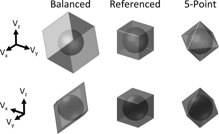
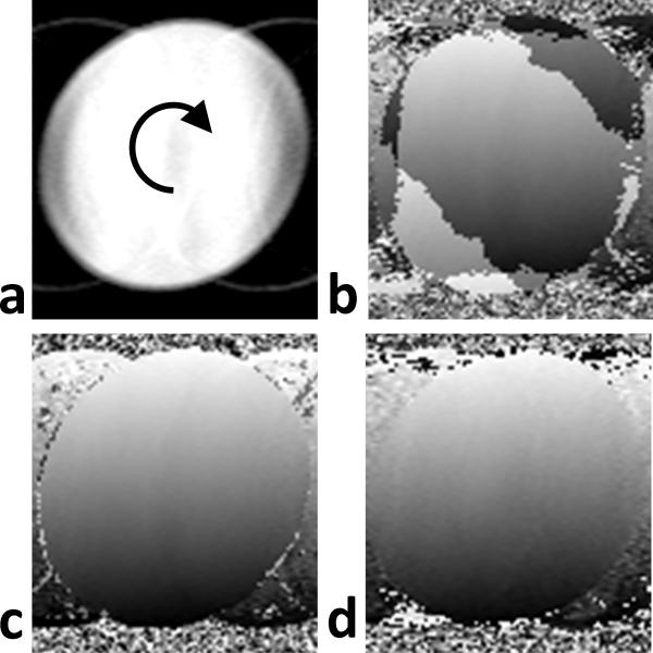
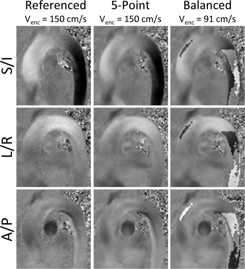
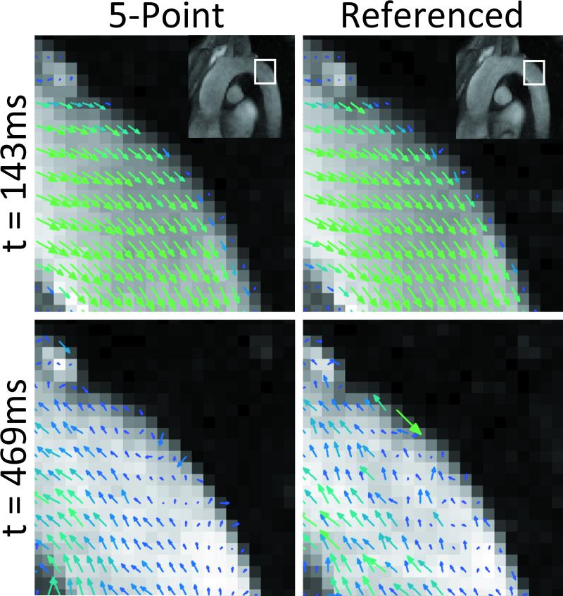
Phase contrast velocimetry can be utilized to measure complex flow for both quantitative and qualitative assessment of vascular hemodynamics. However, phase contrast requires that a maximum measurable velocity be set that balances noise and phase aliasing. To efficiently reduce noise in phase contrast images, several investigators have proposed extended velocity encoding schemes that use extra encodings to unwrap phase aliasing; however, existing techniques can lead to significant increases in echo and scan time, limiting their clinical benefits. In this work, we have developed a novel five-point velocity encoding scheme that efficiently reduces noise with minimal increases in scan and echo time. Investigations were performed in phantoms, demonstrating a 63% increase in velocity-to-noise ratio compared to standard four-point encoding schemes. Aortic velocity measurements were performed in healthy volunteers, showing similar velocity-to-noise ratio improvements. In those volunteers, it was also demonstrated that, without sacrificing accuracy, low-resolution images can be used for the fifth encoding point, reducing the scan time penalty from 25% down to less than 1%.
Figures
References
-
- Markl M, Harloff A, Bley TA, Zaitsev M, Jung B, Weigang E, Langer M, Hennig J, Frydrychowicz A. Time-resolved 3D MR velocity mapping at 3T: improved navigator-gated assessment of vascular anatomy and blood flow. J Magn Reson Imaging. 2007;25(4):824–831. - PubMed
-
- Kvitting JP, Ebbers T, Wigstrom L, Engvall J, Olin CL, Bolger AF. Flow patterns in the aortic root and the aorta studied with time-resolved, 3-dimensional, phase-contrast magnetic resonance imaging: implications for aortic valve-sparing surgery. J Thorac Cardiovasc Surg. 2004;127(6):1602–1607. - PubMed
-
- Wigstrom L, Ebbers T, Fyrenius A, Karlsson M, Engvall J, Wranne B, Bolger AF. Particle trace visualization of intracardiac flow using time-resolved 3D phase contrast MRI. Magn Reson Med. 1999;41(4):793–799. - PubMed
-
- Stalder AF, Russe MF, Frydrychowicz A, Bock J, Hennig J, Markl M. Quantitative 2D and 3D phase contrast MRI: Optimized analysis of blood flow and vessel wall parameters. Magn Reson Med. 2008;60(5):1218–1231. - PubMed
-
- Wu SP, Ringgaard S, Pedersen EM. Three-dimensional phase contrast velocity mapping acquisition improves wall shear stress estimation in vivo. Magn Reson Imaging. 2004;22(3):345–351. - PubMed
Publication types
MeSH terms
Substances
Grants and funding
LinkOut - more resources
Full Text Sources
Other Literature Sources
