

Improved blood suppression in three-dimensional (3D) fast spin-echo (FSE) vessel wall imaging using a combination of double inversion-recovery (DIR) and diffusion sensitizing gradient (DSG) preparations
- PMID: 20099353
- PMCID: PMC6570529
- DOI: 10.1002/jmri.22042
Improved blood suppression in three-dimensional (3D) fast spin-echo (FSE) vessel wall imaging using a combination of double inversion-recovery (DIR) and diffusion sensitizing gradient (DSG) preparations
Abstract
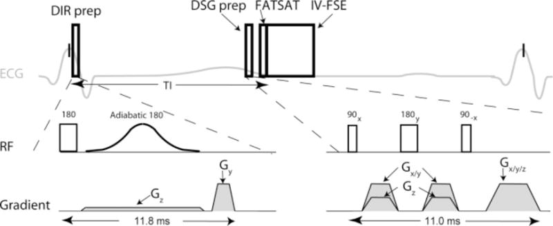
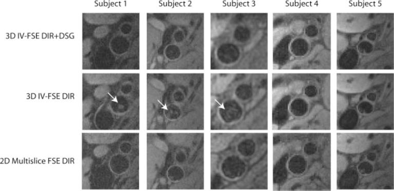
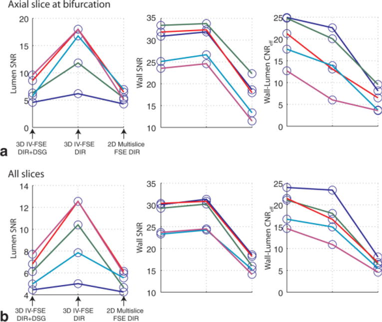
Purpose: To provide improved blood suppression in three-dimensional inner-volume fast spin-echo (3D IV-FSE) carotid vessel wall imaging by using a hybrid preparation consisting of double inversion-recovery (DIR) and diffusion sensitizing gradients (DSG).
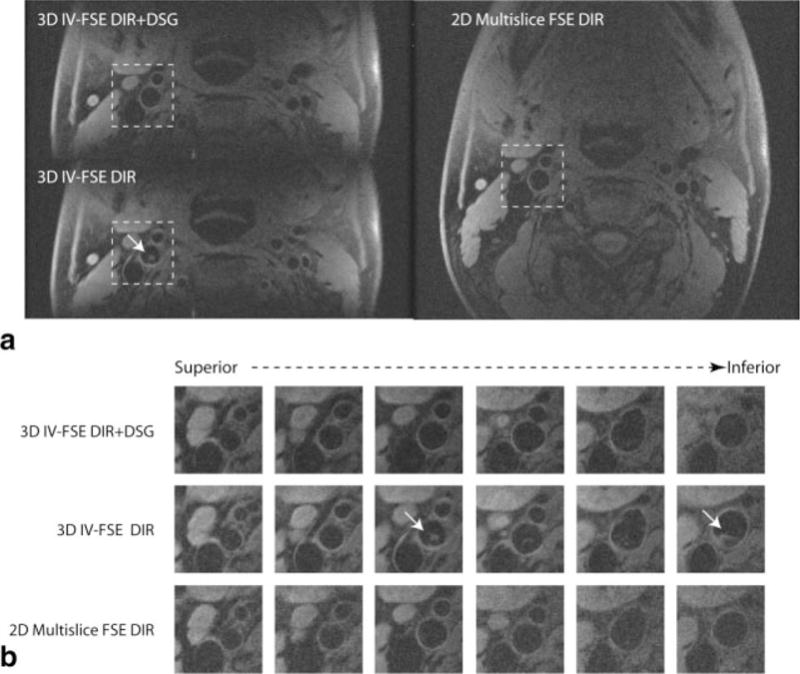
Materials and methods: Multicontrast black-blood MRI is widely used for vessel wall imaging and characterization of atherosclerotic plaque composition. Blood suppression is difficult when using 3D volumetric imaging techniques. DIR approaches do not provide robust blood suppression due to incomplete replacement of blood spins, and DSG approaches compromise vessel wall signal, reducing the lumen-wall contrast-to-noise ratio efficiency (CNR(eff)). In this work a hybrid DIR+DSG preparation is developed and optimized for blood suppression, vessel wall signal preservation, and vessel-wall contrast in 3D IV-FSE imaging. Cardiac gated T(1)-weighted carotid vessel wall images were acquired in five volunteers with 0.5 x 0.5 x 2.5 mm(3) spatial resolution in 80 seconds.
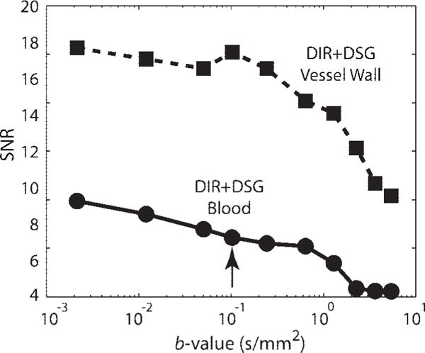
Results: Data from healthy volunteers indicate that the proposed method yields a statistically significant (P < 0.01) improvement in blood suppression and lumen-wall CNR(eff) compared to standard DIR and standard DSG methods alone.
Conclusion: A combination of DIR and DSG preparations can provide improved blood suppression and lumen-wall CNR(eff) for 3D IV-FSE vessel wall imaging.
Figures
Similar articles
-
Three-dimensional flow-independent balanced steady-state free precession vessel wall MRI of the popliteal artery: preliminary experience and comparison with flow-dependent black-blood techniques.J Magn Reson Imaging. 2011 Sep;34(3):696-701. doi: 10.1002/jmri.22663. Epub 2011 Jul 18. J Magn Reson Imaging. 2011. PMID: 21769963 Free PMC article.
-
Relaxation enhanced compressed sensing three-dimensional black-blood vessel wall MR imaging: Preliminary studies.Magn Reson Imaging. 2015 Sep;33(7):932-8. doi: 10.1016/j.mri.2015.03.009. Epub 2015 Apr 8. Magn Reson Imaging. 2015. PMID: 25863136
-
Phase-sensitive dual-inversion recovery for accelerated carotid vessel wall imaging.Invest Radiol. 2015 Mar;50(3):135-43. doi: 10.1097/RLI.0000000000000110. Invest Radiol. 2015. PMID: 25369853 Clinical Trial.
-
Optimization of improved motion-sensitized driven-equilibrium (iMSDE) blood suppression for carotid artery wall imaging.J Cardiovasc Magn Reson. 2014 Aug 9;16(1):61. doi: 10.1186/s12968-014-0061-5. J Cardiovasc Magn Reson. 2014. PMID: 25160911 Free PMC article.
-
Advanced techniques for MRI of atherosclerotic plaque.Top Magn Reson Imaging. 2009 Aug;20(4):217-25. doi: 10.1097/RMR.0b013e3181ea2853. Top Magn Reson Imaging. 2009. PMID: 20805732 Free PMC article. Review.
Cited by
-
Reduction of motion artifacts in carotid MRI using free-induction decay navigators.J Magn Reson Imaging. 2014 Jul;40(1):214-20. doi: 10.1002/jmri.24389. Epub 2013 Nov 13. J Magn Reson Imaging. 2014. PMID: 24677562 Free PMC article.
-
Three-dimensional flow-independent balanced steady-state free precession vessel wall MRI of the popliteal artery: preliminary experience and comparison with flow-dependent black-blood techniques.J Magn Reson Imaging. 2011 Sep;34(3):696-701. doi: 10.1002/jmri.22663. Epub 2011 Jul 18. J Magn Reson Imaging. 2011. PMID: 21769963 Free PMC article.
-
Segmentation of carotid plaque using multicontrast 3D gradient echo MRI.J Magn Reson Imaging. 2012 Apr;35(4):812-9. doi: 10.1002/jmri.22886. Epub 2011 Nov 29. J Magn Reson Imaging. 2012. PMID: 22127812 Free PMC article.
-
High-resolution MR vessel wall imaging in determining the stroke aetiology and risk stratification in isolated middle cerebral artery disease.Neuroradiology. 2022 Aug;64(8):1569-1577. doi: 10.1007/s00234-021-02891-9. Epub 2022 Feb 3. Neuroradiology. 2022. PMID: 35112218
-
Symptomatic unruptured isolated middle cerebral artery dissection: clinical and magnetic resonance imaging features.Clin Neuroradiol. 2016 Mar;26(1):81-91. doi: 10.1007/s00062-014-0337-z. Epub 2014 Sep 6. Clin Neuroradiol. 2016. PMID: 25192774
References
-
- Toussaint JF, LaMuraglia GM, Southern JF, Fuster V, Kantor HL. Magnetic resonance images lipid, fibrous, calcified, hemorrhagic, and thrombotic components of human atherosclerosis in vivo. Circulation. 1996;94:932–938. - PubMed
-
- Soila K, Nummi P, Ekfors T, Viamonte M, Kormano M. Proton relaxation times in arterial wall and atheromatous lesions in man. Invest Radiol. 1986;21:411–415. - PubMed
-
- Shinnar M, Fallon JT, Wehrli S, et al. The diagnostic accuracy of ex vivo MRI for human atherosclerotic plaque characterization. Arterioscler Thromb Vasc Biol. 1999;19:2756–2761. - PubMed
-
- Yuan C, Mitsumori LM, Ferguson MS, et al. In vivo accuracy of multispectral magnetic resonance imaging for identifying lipid-rich necrotic cores and intraplaque hemorrhage in advanced human carotid plaques. Circulation. 2001;104:2051–2056. - PubMed
-
- Koktzoglou I, Chung Y, Mani V, et al. Multislice dark-blood carotid artery wall imaging: a 1.5 T and 3.0 T comparison. J Magn Reson Imaging. 2006;23:699–705. - PubMed
Publication types
MeSH terms
Grants and funding
LinkOut - more resources
Full Text Sources
Other Literature Sources
Medical
