. 2010 Feb 16;49(6):1226-35.
doi: 10.1021/bi901812p.

Structure determination and characterization of the vitamin B6 degradative enzyme (E)-2-(acetamidomethylene)succinate hydrolase

Affiliations

Structure determination and characterization of the vitamin B6 degradative enzyme (E)-2-(acetamidomethylene)succinate hydrolase

Kathryn M McCulloch et al. Biochemistry. .

Abstract

The gene identification and kinetic characterization of (E)-2-(acetamidomethylene)succinate (E-2AMS) hydrolase has recently been described. This enzyme catalyzes the final reaction in the degradation of vitamin B(6) and produces succinic semialdehyde, acetate, ammonia, and carbon dioxide from E-2AMS. The structure of E-2AMS hydrolase was determined to 2.3 A using SAD phasing. E-2AMS hydrolase is a member of the alpha/beta hydrolase superfamily and utilizes a serine/histidine/aspartic acid catalytic triad. Mutation of either the nucleophilic serine or the aspartate resulted in inactive enzyme. Mutation of an additional serine residue in the active site causes the enzyme to be unstable and is likely structurally important. The structure also provides insight into the mechanism of hydrolysis of E-2AMS and identifies several potential catalytically important residues.


Figures

Figure 1
Figure 1
Vitamin B6 degradative pathway found in M. loti. 1, pyridoxine; 2, pyridoxal; 3, 4-pyridoxolactone; 4, 4-pyridoxic acid; 5, 5-formyl-2-methyl-3-hydroxypyridine-4-carboxylic acid; 6, 2-methyl-3-hydroxypyridine-4,5-dicarboxylic acid; 7, 2-methyl-3-hydroxypyridine-5-carboxylic acid; 8, 2-(acetamidomethylene)succinic acid; 9, succinic semialdehyde. Other final products are ammonia, acetate, and carbon dioxide.
Figure 2
Figure 2
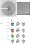
Pseudo-translational symmetry in E-2AMS hydrolase crystals. A. A typical diffraction pattern for E-2AMS hydrolase. At low resolution, every third row of reflections is much more intense than other rows. B. Crystal packing of E-2AMS hydrolase. For clarity only one third of the unit cell is shown, with the three unique chains colored blue, red and green. The crystallographically related protomers are shown in grey.
Figure 3
Figure 3
Monomeric structure of E-2AMS hydrolase. A. Stereoview ribbon diagram of E-2AMS hydrolase with the secondary structures labelled. β-strands are shown in green and α-helices are shown in blue. B. Topology diagram of E-2AMS hydrolase, using the same color scheme as A. C. Ribbon diagram of E-2AMS hydrolase illustrating the sharp twist in the β-sheet running through the core of the enzyme and the cap domain.
Figure 4
Figure 4
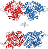
Dimeric structure of E-2AMS hydrolase. Each chain is colored differently, one red and the second blue. The second orientation obtained by rotating the first orientation by 90° along the horizontal axis.
Figure 5
Figure 5
Stereoview ball and stick diagram of the active site of E-2AMS hydrolase. Water molecules are shown as red nonbonded spheres and the chloride ion is shown as a nonbonded grey sphere. Key protein interactions are shown as dashed lines.
Figure 6
Figure 6
Sequence alignment of E-2AMS hydrolase with other enzymes identified as structurally homologous by DALI. Included enzymes are the α/β hydrolase YP_496220.1 (3BWX), CarC (1J1I), chloroperoxidase L (1A88), the PFE arylesterase (1VA4), the EST esterase (1ZOI), ybfF (3BF7), fluoroacetate dehalogenase (1Y37), and MhpC (1U2E). The nucleophile is marked with a green circle, the catalytic histidine is labelled with a blue star, and the different active site acids are labelled using triangles. The expected acidic residues have a purple triangle, while the location of the acidic residue in E-2AMS hydrolase has a yellow triangle.
Figure 7
Figure 7
Stereoview ball and stick diagram comparing the active site of E-2AMS hydrolase to other members of the α/β hydrolase superfamily. The E-2AMS hydrolase structure is shown with green carbon atoms, ybfF is shown using magenta carbon atoms, and the carbon-carbon bond hydrolase MhpC has white carbon atoms. All residues of E-2AMS hydrolase are labelled and the catalytic residues of ybfF and MhpC are labelled.
Figure 8
Figure 8
Proposed mechanisms for the hydrolysis of E-2AMS to produce succinic semialdehyde, acetate, ammonia, and carbon dioxide. Mechanism 1 utilizes Ser106 to activate a water molecule for attack at the carbonyl carbon of the amide bond, while Mechanism 2 utilizes a direct attack on E-2AMS by Ser106.
Figure 9
Figure 9
Possible coordination of E-2AMS within the active site. A. Schematic diagram where E-2AMS (shown in red) was manually positioned such that either Ser106 or an activated water molecule could attack the carbonyl carbon of the amide group. Potential hydrogen bonds are shown as dashed lines. B. Stereoview ball and stick diagram of the E-2AMS hydrolase active site with E-2AMS manually positioned in the active site. Carbon atoms of E-2AMS hydrolase are colored green and carbon atoms of E-2AMS are colored gray. Possible interactions and hydrogen bonds important for active site orientation are shown in dashed lines.

Similar articles

Cited by

References

    1. Eliot AC, Kirsch JF. Pyridoxal phosphate enzymes: mechanistic, structural, and evolutionary considerations. Annu. Rev. Biochem. 2004;73:383–415. - PubMed
    1. Snell EE, Haskell BE. The Metabolism of Vitamin B6, In: Comprehensive Biochemistry. Vol. 21. New York: Elsevier/North Holland; 1971.
    1. Kaneko T, Nakamura Y, Sato S, Asamizu E, Kato T, Sasamoto S, Watanabe A, Idesawa K, Ishikawa A, Kawashima K, Kimura T, Kishida Y, Kiyokawa C, Kohara M, Matsumoto M, Matsuno A, Mochizuki Y, Nakayama S, Nakazaki N, Shimpo S, Sugimoto M, Takeuchi C, Yamada M, Tabata S. Complete genome structure of the nitrogen-fixing symbiotic bacterium Mesorhizobium loti. DNA Res. 2000;7:331–338. - PubMed
    1. Yuan B, Yoshikane Y, Yokochi N, Ohnishi K, Yagi T. The nitrogen-fixing symbiotic bacterium Mesorhizobium loti has and expresses the gene encoding pyridoxine 4-oxidase involved in the degradation of vitamin B6. FEMS Microbiol. Lett. 2004;234:225–230. - PubMed
    1. Funami J, Yoshikane Y, Kobayashi H, Yokochi N, Yuan B, Iwasaki K, Ohnishi K, Yagi T. 4-Pyridoxolactonase from a symbiotic nitrogen-fixing bacterium Mesorhizobium loti: cloning, expression, and characterization. Biochim. Biophys. Acta. 2005;1753:234–239. - PubMed

Publication types

Associated data

LinkOut - more resources