

Combining chemoselective ligation with polyhistidine-driven self-assembly for the modular display of biomolecules on quantum dots
- PMID: 20099912
- PMCID: PMC4756922
- DOI: 10.1021/nn901393v
Combining chemoselective ligation with polyhistidine-driven self-assembly for the modular display of biomolecules on quantum dots
Abstract
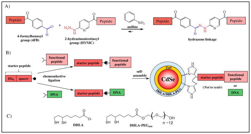
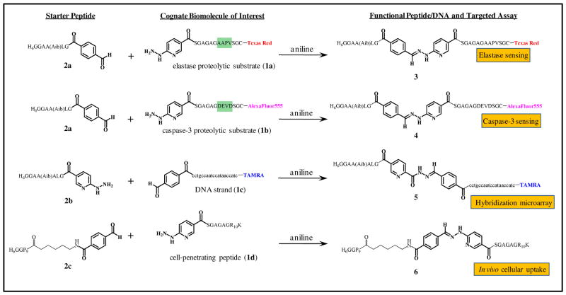
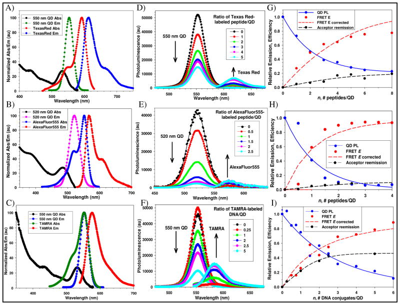
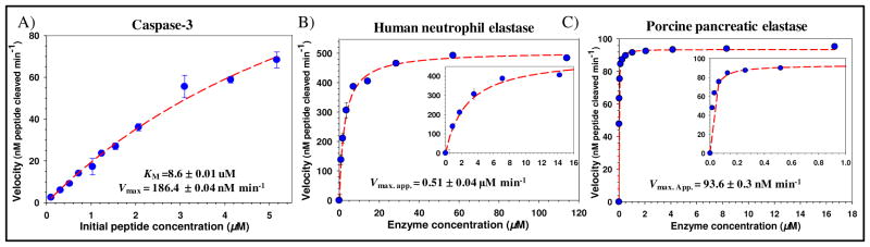
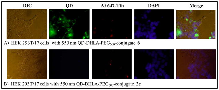
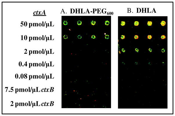
One of the principle hurdles to wider incorporation of semiconductor quantum dots (QDs) in biology is the lack of facile linkage chemistries to create different types of functional QD--bioconjugates. A two-step modular strategy for the presentation of biomolecules on CdSe/ZnS core/shell QDs is described here which utilizes a chemoselective, aniline-catalyzed hydrazone coupling chemistry to append hexahistidine sequences onto peptides and DNA. This specifically provides them the ability to ratiometrically self-assemble to hydrophilic QDs. The versatility of this labeling approach was highlighted by ligating proteolytic substrate peptides, an oligoarginine cell-penetrating peptide, or a DNA-probe to cognate hexahistidine peptidyl sequences. The modularity allowed subsequently self-assembled QD constructs to engage in different types of targeted bioassays. The self-assembly and photophysical properties of individual QD conjugates were first confirmed by gel electrophoresis and Forster resonance energy transfer analysis. QD-dye-labeled peptide conjugates were then used as biosensors to quantitatively monitor the proteolytic activity of caspase-3 or elastase enzymes from different species. These sensors allowed the determination of the corresponding kinetic parameters, including the Michaelis constant (K(M)) and the maximum proteolytic activity (V(max)). QDs decorated with cell-penetrating peptides were shown to be successfully internalized by HEK 293T/17 cells, while nanocrystals displaying peptide--DNA conjugates were utilized as fluorescent probes in hybridization microarray assays. This modular approach for displaying peptides or DNA on QDs may be extended to other more complex biomolecules such as proteins or utilized with different types of nanoparticle materials.
Figures
Similar articles
-
Quantum dot peptide biosensors for monitoring caspase 3 proteolysis and calcium ions.ACS Nano. 2010 Sep 28;4(9):5487-97. doi: 10.1021/nn1016132. ACS Nano. 2010. PMID: 20822159
-
Synthesizing and modifying peptides for chemoselective ligation and assembly into quantum dot-peptide bioconjugates.Methods Mol Biol. 2013;1025:47-73. doi: 10.1007/978-1-62703-462-3_5. Methods Mol Biol. 2013. PMID: 23918329
-
A reactive peptidic linker for self-assembling hybrid quantum dot-DNA bioconjugates.Nano Lett. 2007 Jun;7(6):1741-8. doi: 10.1021/nl070782v. Epub 2007 May 26. Nano Lett. 2007. PMID: 17530814
-
Quantum Dot-Dye Conjugates for Biosensing, Imaging, and Therapy.Adv Healthc Mater. 2018 Jul;7(14):e1800252. doi: 10.1002/adhm.201800252. Epub 2018 Jun 3. Adv Healthc Mater. 2018. PMID: 29862653 Free PMC article. Review.
-
Next-Generation DNA-Functionalized Quantum Dots as Biological Sensors.ACS Chem Biol. 2018 Jul 20;13(7):1705-1713. doi: 10.1021/acschembio.7b00887. Epub 2018 Jan 4. ACS Chem Biol. 2018. PMID: 29257662 Review.
Cited by
-
QD-Based FRET Probes at a Glance.Sensors (Basel). 2015 Jun 4;15(6):13028-51. doi: 10.3390/s150613028. Sensors (Basel). 2015. PMID: 26053750 Free PMC article. Review.
-
Aromatic aldehyde and hydrazine activated peptide coated quantum dots for easy bioconjugation and live cell imaging.Bioconjug Chem. 2011 Jun 15;22(6):1006-11. doi: 10.1021/bc100593m. Epub 2011 May 16. Bioconjug Chem. 2011. PMID: 21553893 Free PMC article.
-
Quantum dot-mediated delivery of siRNA to inhibit sphingomyelinase activities in brain-derived cells.J Neurochem. 2016 Dec;139(5):872-885. doi: 10.1111/jnc.13841. Epub 2016 Oct 14. J Neurochem. 2016. PMID: 27622309 Free PMC article.
-
Proteintemplat-gesteuerte Fragmentligationen - von der molekularen Erkennung zur Wirkstofffindung.Angew Chem Weinheim Bergstr Ger. 2017 Jun 19;129(26):7464-7485. doi: 10.1002/ange.201610372. Epub 2017 May 31. Angew Chem Weinheim Bergstr Ger. 2017. PMID: 32313319 Free PMC article.
-
Robust and specific ratiometric biosensing using a copper-free clicked quantum dot-DNA aptamer sensor.Nanoscale. 2013 Nov 7;5(21):10307-15. doi: 10.1039/c3nr02897f. Epub 2013 Sep 11. Nanoscale. 2013. PMID: 24056667 Free PMC article.
References
-
- Klostranec JM, Chan WCW. Quantum Dots in Biological and Biomedical Research: Recent Progress and Present Challenges. Advanced Materials. 2006;18:1953–1964.
-
- Algar WR, Massey M, Krull UJ. The Application of Quantum Dots, Gold Nanoparticles and Molecular Switches to Optical Nucleic-Acid Diagnostics. Trac-Trends in Analytical Chemistry. 2009;28:292–306.
-
- Goldman ER, Clapp AR, Anderson GP, Uyeda HT, Mauro JM, Medintz IL, Mattoussi H. Multiplexed Toxin Analysis Using Four Colors of Quantum Dot Fluororeagents. Analyical Chemistry. 2004;76:684–688. - PubMed
-
- Medintz IL, Farrell D, Susumu K, Trammell SA, Deschamps JR, Brunel FM, Dawson PE, Mattoussi HM. Multiplex Charge Transfer Interactions Between Quantum Dots and Peptide-Bridged Ruthenium Complexes. Analyical Chemistry. 2009;81:4831–4839. - PubMed
Publication types
MeSH terms
Substances
Grants and funding
LinkOut - more resources
Full Text Sources
Research Materials
Miscellaneous
