. 2010 Jun;16(6):2017-27.
doi: 10.1089/ten.TEA.2009.0768.

Design and characterization of an injectable pericardial matrix gel: a potentially autologous scaffold for cardiac tissue engineering

Affiliations

Design and characterization of an injectable pericardial matrix gel: a potentially autologous scaffold for cardiac tissue engineering

Sonya B Seif-Naraghi et al. Tissue Eng Part A. 2010 Jun.

Abstract

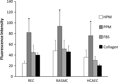
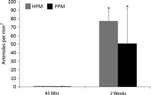
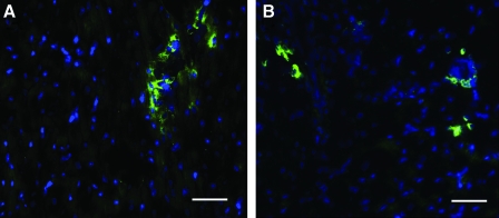
Following ischemic injury in the heart, little to no repair occurs, causing a progressive degeneration of cardiac function that leads to congestive heart failure. Cardiac tissue engineering strategies have focused on designing a variety of injectable scaffolds that range in composition from single-component materials to complex extracellular matrix (ECM)-derived materials. In this study, the pericardial ECM, a commonly used biomaterial, was investigated for use as an injectable scaffold for cardiac repair. It was determined that a solubilized form of decellularized porcine pericardium could be injected and induced to gel in vivo, prompting investigation with human pericardium, which has the decided advantage of offering an autologous therapy. Characterization showed that the matrix gels retained components of the native pericardial ECM, with extant protein and glycosaminoglycan content identified. The results of an in vitro migration assay indicate that the porcine pericardial matrix is a stronger chemoattractant for relevant cell types, but in vivo results showed that the two materials caused statistically similar amounts of neovascularization, demonstrating feasibility as injectable treatments. Potential stem cell mobilization was supported by the presence of c-Kit+ cells within the matrix injection regions. With this work, the pericardium is identified as a novel source for an autologous scaffold for treating myocardial infarction.


Figures

FIG. 1.
FIG. 1.
Histological analysis of pericardia. Hematoxylin and eosin (H&E) stains of (A) fresh human pericardium, (B) decellularized human pericardial matrix, (C) fresh porcine pericardium, and (D) decellularized porcine pericardial matrix. Scale bars: 500 μm. Color images available online at www.liebertonline.com/ten.
FIG. 2.
FIG. 2.
Tissue processing. Decellularized porcine pericardium (A) was lyophilized (B), milled into a powder (C), and then solubilized (D). Color images available online at www.liebertonline.com/ten.
FIG. 3.
FIG. 3.
Polyacrylamide gel electrophoresis results. (A) Molecular weight standard. (B) Rat tail collagen type I (2.5 mg/mL) compared to the solubilized human (C) and porcine (D) pericardial extracellular matrix (7 mg/mL). Note the presence of collagen as well as several other proteins/peptides in the pericardial matrix samples. Color images available online at www.liebertonline.com/ten.
FIG. 4.
FIG. 4.
In vitro migration assay. REC, RASMC, and HCAEC migration toward solubilized human pericardial matrix (HPM), porcine pericardial matrix (PPM), collagen, and 10% fetal bovine serum. Values are based on fluorescence data–intensity correlates to the number of cells that have migrated through the transwell membrane toward any given solution. *p < 0.05 compared to other groups. REC, rat epicardial cell; RASMC, rat aortic smooth muscle cell; HCAEC, human coronary artery endothelial cell.
FIG. 5.
FIG. 5.
Myocardial injections: in vivo gelation. H&E stain of human (A) and porcine (B) pericardial matrix injections that have gelled in vivo after 45 min. Arrows denote matrix location, stained lighter pink than the myocardium. Scale bars: 500 μm. Color images available online at www.liebertonline.com/ten.
FIG. 6.
FIG. 6.
Vasculature infiltration. Fluorescent stains for vessels in the injected human (A–C) and porcine (E–G) matrix gels at 2 weeks. Endothelial cells are labeled green (A, E), whereas smooth muscle cells are labeled red (B, F). Merged images are shown in (C) and (G). Scale bars: 100 μm. The white or black dotted lines indicate the area of matrix injection, as determined by H&E analysis of a nearby section for both human (D) and porcine (H) matrix injections. Color images available online at www.liebertonline.com/ten.
FIG. 7.
FIG. 7.
Quantification of arteriole density in matrix injection regions. Injection of human and porcine pericardial matrix caused a neovascularization response that was quantified by the increase in the average number of vessels with average diameters greater than 10 μm per mm2 of injection region after 2 weeks compared to 45 min (*p < 0.01). The two materials caused a statistically similar response (p = 0.26).
FIG. 8.
FIG. 8.
Stem cells within matrix injection region. A Hoechst stain for nuclei (blue) and an immunohistochemical stain for c-kit (green) identifies stem cells in the human (A) and porcine (B) matrix injection regions. Scale bars: 50 μm. Color images available online at www.liebertonline.com/ten.

References

    1. Lloyd-Jones D. Adams R. Carnethon M. De Simone G. Ferguson T.B. Flegal K. Ford E. Furie K. Go A. Greenlund K. Haase N. Hailpern S. Ho M. Howard V. Kissela B. Kittner S. Lackland D. Lisabeth L. Marelli A. McDermott M. Meigs J. Mozaffarian D. Nichol G. O'Donnell C. Roger V. Rosamond W. Sacco R. Sorlie P. Stafford R. Steinberger J. Thom T. Wasserthiel-Smoller S. Wong N. Wylie-Rosett J. Hong Y. American Heart Association Statistics Committee and Stroke Statistics Subcommittee. Heart Disease and Stroke Statistics—2009 Update: A report from the American Heart Association Statistics Committee and Stroke Statistics Subcommittee. Circulation. 2009;119:e21. - PubMed
    1. Bergmann O. Bhardwaj R.D. Bernard S. Zdunek S. Barnabe-Heider F. Walsh S. Zupicich J. Alkass K. Buchholz B.A. Druid H. Jovinge S. Frisen J. Evidence for cardiomyocyte renewal in humans. Science. 2009;324:98. - PMC - PubMed
    1. Mann D.L. Mechanisms and models in heart failure: a combinatorial approach. Circulation. 1999;100:999. - PubMed
    1. Laflamme M.A. Murry C.E. Regenerating the heart. Nat Biotechnol. 2005;23:845. - PubMed
    1. Reffelmann T. Kloner R.A. Cellular cardiomyoplasty—cardiomyocytes, skeletal myoblasts, or stem cells for regenerating myocardium and treatment of heart failure? Cardiovasc Res. 2003;58:358. - PubMed

Publication types

LinkOut - more resources