

Alopecia in IL-10-deficient mouse pups is c-kit-dependent and can be triggered by iron deficiency
- PMID: 20100190
- PMCID: PMC3514968
- DOI: 10.1111/j.1600-0625.2009.01032.x
Alopecia in IL-10-deficient mouse pups is c-kit-dependent and can be triggered by iron deficiency
Abstract
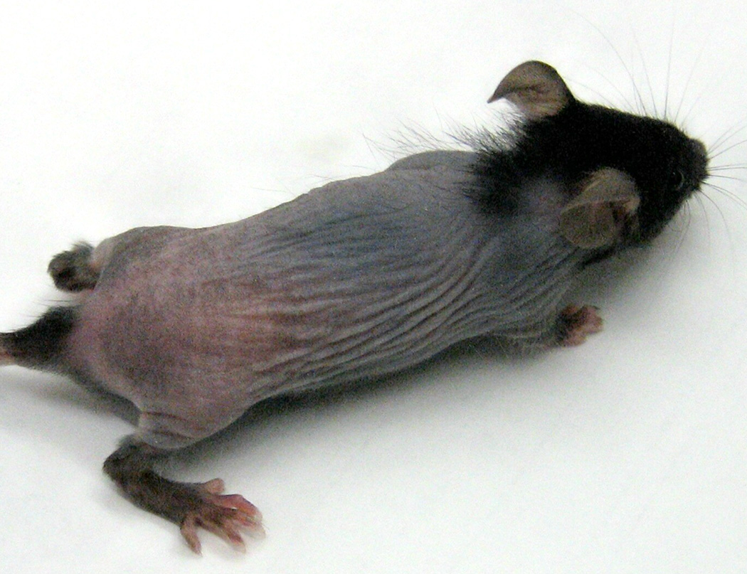
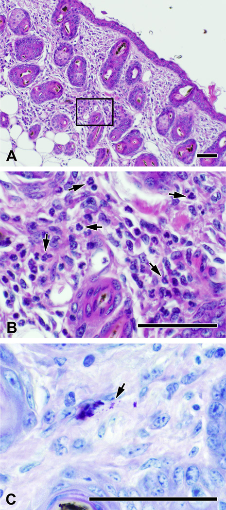
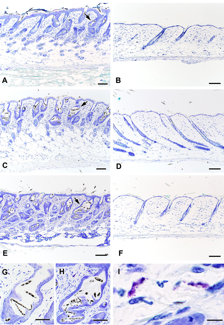
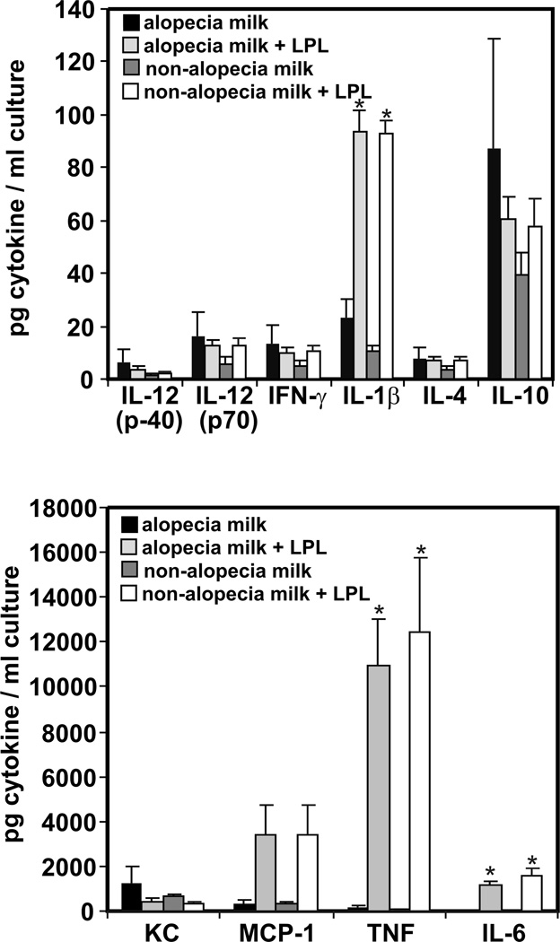
Hair loss (alopecia) can result from a variety of metabolic, endocrine, immunologic, and environmental causes. This investigation was undertaken to determine the mechanisms underlying the sporadic development of alopecia in litters from C57BL/6 interleukin-10-deficient (Il10(-/-)) mice. All pups in affected litters demonstrated alopecia by postnatal days 17-19, with hair loss from their trunks but not from their head, base of tail, or feet. Histopathology revealed distorted hair follicles containing broken hair shafts and prominent dermal infiltrates containing increased numbers of activated mast cells. Hair re-growth began soon after weaning, suggesting that the alopecia was triggered by factors transmitted during lactation. Milk from Il10(-/-) dams induced macrophage secretion of pro-inflammatory cytokines in vitro regardless of whether or not their pups developed alopecia. Feeding dams a diet containing 3-6 ppm iron increased the percentage of litters with alopecia to 100% for pups with mast cells, with 0% alopecia in mast cell-deficient pups. When dams were fed a diet containing 131 ppm iron, significantly lower haemoglobin and hematocrit values were observed in pups from litters with alopecia (71%; 5 of 7 litters) compared to litters without alopecia. Genetic or pharmacologic inhibition of c-kit that resulted in depletion of mast cells in pups prevented hair loss in at-risk litters. These studies demonstrate that maternal iron-restricted diets enhance the incidence of alopecia in IL-10-deficient mouse pups and suggest mast cells as potential effector cells. Further studies are indicated to further explore the mechanisms involved and to determine how mast cells may contribute to alopecia in humans.
Figures

Similar articles
-
NTP technical report on the toxicity studies of Dibutyl Phthalate (CAS No. 84-74-2) Administered in Feed to F344/N Rats and B6C3F1 Mice.Toxic Rep Ser. 1995 Apr;30:1-G5. Toxic Rep Ser. 1995. PMID: 12209194
-
Pregnancy in postpartum estrus induces inflammatory milk production and catagen specific pup skin inflammation in interleukin-10 deficient mice.J Dermatol Sci. 2013 Dec;72(3):225-32. doi: 10.1016/j.jdermsci.2013.07.004. Epub 2013 Jul 23. J Dermatol Sci. 2013. PMID: 23928228
-
Impaired natural killer cell activity in iron-deficient rat pups.J Nutr. 1987 Mar;117(3):567-71. doi: 10.1093/jn/117.3.567. J Nutr. 1987. PMID: 3572568
-
Stem cells and alopecia: a review of pathogenesis.Br J Dermatol. 2012 Sep;167(3):479-84. doi: 10.1111/j.1365-2133.2012.11018.x. Epub 2012 Aug 8. Br J Dermatol. 2012. PMID: 22533551 Review.
-
The diagnosis and treatment of iron deficiency and its potential relationship to hair loss.J Am Acad Dermatol. 2006 May;54(5):824-44. doi: 10.1016/j.jaad.2005.11.1104. J Am Acad Dermatol. 2006. PMID: 16635664 Review.
Cited by
-
Iron supplementation decreases severity of allergic inflammation in murine lung.PLoS One. 2012;7(9):e45667. doi: 10.1371/journal.pone.0045667. Epub 2012 Sep 20. PLoS One. 2012. Retraction in: PLoS One. 2016 May 09;11(5):e0155387. doi: 10.1371/journal.pone.0155387. PMID: 23029172 Free PMC article. Retracted.
-
Malnutrition and Allergies: Tipping the Immune Balance towards Health.J Clin Med. 2024 Aug 11;13(16):4713. doi: 10.3390/jcm13164713. J Clin Med. 2024. PMID: 39200855 Free PMC article. Review.
-
Imatinib resistance and microcytic erythrocytosis in a KitV558Δ;T669I/+ gatekeeper-mutant mouse model of gastrointestinal stromal tumor.Proc Natl Acad Sci U S A. 2012 Aug 21;109(34):E2276-83. doi: 10.1073/pnas.1115240109. Epub 2012 May 31. Proc Natl Acad Sci U S A. 2012. PMID: 22652566 Free PMC article.
-
Reproduction and Growth in a Murine Model of Early Life-Onset Inflammatory Bowel Disease.PLoS One. 2016 Apr 5;11(4):e0152764. doi: 10.1371/journal.pone.0152764. eCollection 2016. PLoS One. 2016. PMID: 27045690 Free PMC article.
-
Why Serological Responses during Cystitis are Limited.Pathogens. 2016 Feb 14;5(1):19. doi: 10.3390/pathogens5010019. Pathogens. 2016. PMID: 26907352 Free PMC article. Review.
References
-
- Paus R, Muller-Rover S, van der Veen C, Maurer M, et al. A Comprehensive Guide for the Recognition and Classification of Distinct Stages of Hair Follicle Morphogenesis. J Invest Dermatol. 1999;113:523–532. - PubMed
-
- Sundberg JP, editor. Handbook of Mouse Mutations with Skin and Hair Abnormalities: Animal Models and Biomedical Tools. Boca Raton: CRC Press; 1994. p. 544.
-
- Mitsiadis TA, Barrandon O, Rochat A, Barrandon Y, De Bari C. Stem cell niches in mammals. Exp Cell Res. 2007;313:3377–3385. - PubMed
-
- Paus R, Cotsarelis G. The biology of hair follicles. N Engl J Med. 1999;341:491–497. - PubMed
-
- Stenn KS, Paus R. Controls of Hair Follicle Cycling. Physiol Rev. 2001;81:449–494. - PubMed
Publication types
MeSH terms
Substances
Grants and funding
LinkOut - more resources
Full Text Sources
