

A role for NRAGE in NF-kappaB activation through the non-canonical BMP pathway
- PMID: 20100315
- PMCID: PMC2829509
- DOI: 10.1186/1741-7007-8-7
A role for NRAGE in NF-kappaB activation through the non-canonical BMP pathway
Abstract
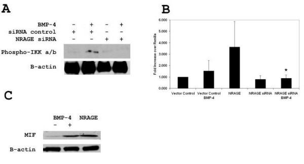
Background: Previous studies have linked neurotrophin receptor-interacting MAGE protein to the bone morphogenic protein signaling pathway and its effect on p38 mediated apoptosis of neural progenitor cells via the XIAP-Tak1-Tab1 complex. Its effect on NF-kappaB has yet to be explored.
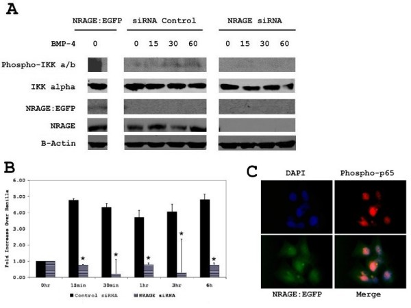
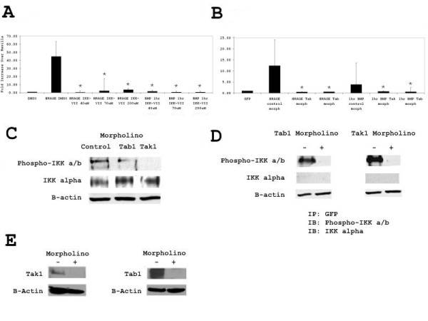
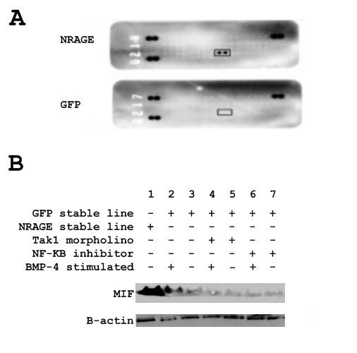
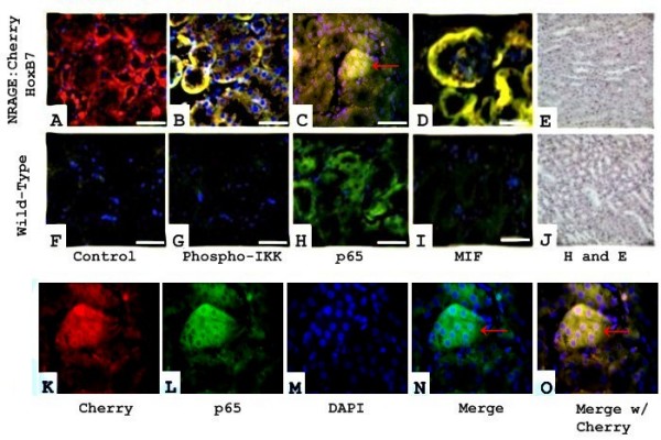
Results: Herein we report that NRAGE, via the same XIAP-Tak1-Tab1 complex, is required for the phosphorylation of IKK -alpha/beta and subsequent transcriptional activation of the p65 subunit of NF-kappaB. Ablation of endogenous NRAGE by siRNA inhibited NF-kappaB pathway activation, while ablation of Tak1 and Tab1 by morpholino inhibited overexpression of NRAGE from activating NF-kappaB. Finally, cytokine profiling of an NRAGE over-expressing stable line revealed the expression of macrophage migration inhibitory factor.
Conclusion: Modulation of NRAGE expression revealed novel roles in regulating NF-kappaB activity in the non-canonical bone morphogenic protein signaling pathway. The expression of macrophage migration inhibitory factor by bone morphogenic protein -4 reveals novel crosstalk between an immune cytokine and a developmental pathway.
Figures
Similar articles
-
The F-box protein FBXO7 positively regulates bone morphogenetic protein-mediated signaling through Lys-63-specific ubiquitination of neurotrophin receptor-interacting MAGE (NRAGE).Cell Mol Life Sci. 2015 Jan;72(1):181-95. doi: 10.1007/s00018-014-1665-5. Epub 2014 Jun 20. Cell Mol Life Sci. 2015. PMID: 24947323 Free PMC article.
-
A small peptide modeled after the NRAGE repeat domain inhibits XIAP-TAB1-TAK1 signaling for NF-κB activation and apoptosis in P19 cells.PLoS One. 2011;6(7):e20659. doi: 10.1371/journal.pone.0020659. Epub 2011 Jul 18. PLoS One. 2011. PMID: 21789165 Free PMC article.
-
Knockdown of NRAGE induces odontogenic differentiation by activating NF-κB signaling in mouse odontoblast-like cells.Connect Tissue Res. 2019 Mar;60(2):71-84. doi: 10.1080/03008207.2018.1439484. Epub 2018 Feb 22. Connect Tissue Res. 2019. PMID: 29448842
-
NRAGE mediates p38 activation and neural progenitor apoptosis via the bone morphogenetic protein signaling cascade.Mol Cell Biol. 2005 Sep;25(17):7711-24. doi: 10.1128/MCB.25.17.7711-7724.2005. Mol Cell Biol. 2005. PMID: 16107717 Free PMC article.
-
NRAGE: a potential rheostat during branching morphogenesis.Mech Dev. 2009 May-Jun;126(5-6):337-49. doi: 10.1016/j.mod.2009.02.005. Epub 2009 Mar 4. Mech Dev. 2009. PMID: 19268530 Free PMC article.
Cited by
-
The F-box protein FBXO7 positively regulates bone morphogenetic protein-mediated signaling through Lys-63-specific ubiquitination of neurotrophin receptor-interacting MAGE (NRAGE).Cell Mol Life Sci. 2015 Jan;72(1):181-95. doi: 10.1007/s00018-014-1665-5. Epub 2014 Jun 20. Cell Mol Life Sci. 2015. PMID: 24947323 Free PMC article.
-
RNA m6A methylation and regulatory proteins in pulmonary arterial hypertension.Hypertens Res. 2024 May;47(5):1273-1287. doi: 10.1038/s41440-024-01607-9. Epub 2024 Mar 4. Hypertens Res. 2024. PMID: 38438725 Review.
-
microRNA-140 Inhibits Inflammation and Stimulates Chondrogenesis in a Model of Interleukin 1β-induced Osteoarthritis.Mol Ther Nucleic Acids. 2016 Oct 11;5(10):e373. doi: 10.1038/mtna.2016.64. Mol Ther Nucleic Acids. 2016. PMID: 27727249 Free PMC article.
-
Cell death in glioblastoma and the central nervous system.Cell Oncol (Dordr). 2025 Apr;48(2):313-349. doi: 10.1007/s13402-024-01007-8. Epub 2024 Nov 6. Cell Oncol (Dordr). 2025. PMID: 39503973 Free PMC article. Review.
-
Anti-Inflammatory Peptide Attenuates Edema and Promotes BMP-2-Induced Bone Formation in Spine Fusion.Tissue Eng Part A. 2018 Nov;24(21-22):1641-1651. doi: 10.1089/ten.TEA.2017.0512. Epub 2018 Jul 3. Tissue Eng Part A. 2018. PMID: 29766758 Free PMC article.
References
-
- Carmody RJ, Chen YH. Nuclear factor-kappaB: activation and regulation during toll-like receptor signaling. Cell Mol Immunol. 2007;4:21–41. - PubMed
Publication types
MeSH terms
Substances
Grants and funding
LinkOut - more resources
Full Text Sources
Miscellaneous
