. 2010 Jan 25:7:3.
doi: 10.1186/1742-4690-7-3.

Cell-specific targeting of lentiviral vectors mediated by fusion proteins derived from Sindbis virus, vesicular stomatitis virus, or avian sarcoma/leukosis virus

Affiliations

Cell-specific targeting of lentiviral vectors mediated by fusion proteins derived from Sindbis virus, vesicular stomatitis virus, or avian sarcoma/leukosis virus

Xian-Yang Zhang et al. Retrovirology. .

Abstract

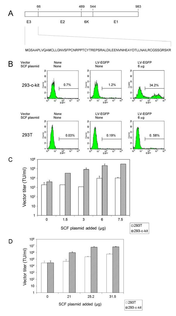
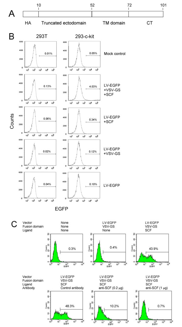
Background: The ability to efficiently and selectively target gene delivery vectors to specific cell types in vitro and in vivo remains one of the formidable challenges in gene therapy. We pursued two different strategies to target lentiviral vector delivery to specific cell types. In one of the strategies, vector particles bearing a membrane-bound stem cell factor sequence plus a separate fusion protein based either on Sindbis virus strain TR339 glycoproteins or the vesicular stomatitis virus G glycoprotein were used to selectively transduce cells expressing the corresponding stem cell factor receptor (c-kit). An alternative approach involved soluble avian sarcoma/leukosis virus receptors fused to cell-specific ligands including stem cell factor and erythropoietin for targeting lentiviral vectors pseudotyped with avian sarcoma/leukosis virus envelope proteins to cells that express the corresponding receptors.

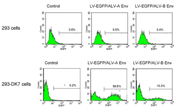
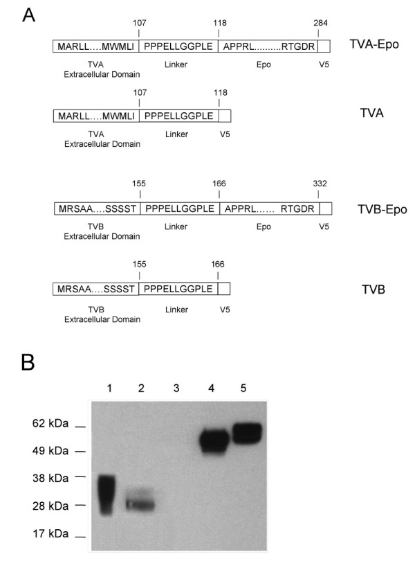
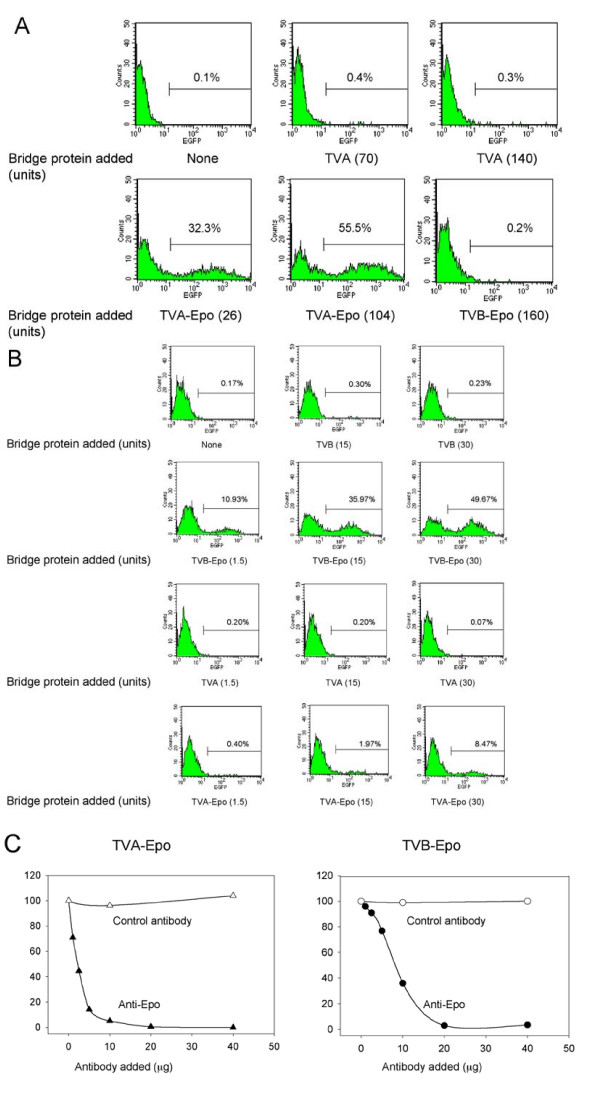
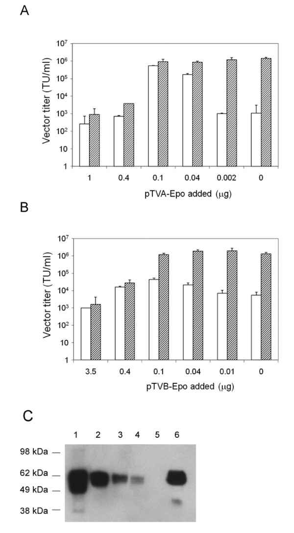
Results: The titers of unconcentrated vector particles bearing Sindbis virus strain TR339 or vesicular stomatitis virus G fusion proteins plus stem cell factor in the context of c-kit expressing cells were up to 3.2 x 10(5) transducing units per ml while vector particles lacking the stem cell factor ligand displayed titers that were approximately 80 fold lower. On cells that lacked the c-kit receptor, the titers of stem cell factor-containing vectors were approximately 40 times lower compared to c-kit-expressing cells. Lentiviral vectors pseudotyped with avian sarcoma/leukosis virus subgroup A or B envelope proteins and bearing bi-functional bridge proteins encoding erythropoietin or stem cell factor fused to the soluble extracellular domains of the avian sarcoma/leukosis virus subgroup A or B receptors resulted in efficient transduction of erythropoietin receptor or c-kit-expressing cells. Transduction of erythropoietin receptor-expressing cells mediated by bi-functional bridge proteins was found to be dependent on the dose, the correct subgroup-specific virus receptor and the correct envelope protein. Furthermore, transduction was completely abolished in the presence of anti-erythropoietin antibody.

Conclusions: Our results indicate that the avian sarcoma/leukosis virus bridge strategy provides a reliable approach for cell-specific lentiviral vector targeting. The background levels were lower compared to alternative strategies involving Sindbis virus strain TR339 or vesicular stomatitis virus fusion proteins.


Figures

Figure 1
Figure 1
Transduction of c-kit-expressing 293 cells using lentiviral vectors pseudotyped with the Sindbis virus strain TR339 glycoproteins and bearing SCF. (A) Schematic representation of the modified Sindbis virus strain TR339 proteins. The sequence of the modified E3 protein is shown. It encodes 66 amino acids including the signal peptide for the E2 protein. E2 consists of 423 amino acids, and the 6K and E1 proteins encode 55 and 439 amino acids, respectively. The numbers refer to the ends of the respective protein domains. (B) Representative FACS profiles. Top panels: 293-c-kit cells transduced with vectors prepared using 0 and 6 μg of the SCF-encoding pUB-HuMGF plasmid; Top left panel: Untransduced cells. Bottom panels: 293T cells transduced with vectors prepared using 0 and 6 μg of the SCF-encoding pUB-HuMGF plasmid; Bottom left panel: Untransduced cells. The percentages of EGFP-positive cells are indicated. (C) Unconcentrated vector titers on 293 cells expressing c-kit (striped bars) and on 293T cells (open bars) three days after transduction. The titers shown represent the mean ± standard deviation (SD) obtained from three independent experiments. (D) Determination of concentrated vector titers. Vectors were concentrated 300 fold by ultracentrifugation. The titers shown represent the mean ± SD from three to six independent experiments. Striped bars: 293 cells expressing c-kit; Open bars: 293T cells.
Figure 2
Figure 2
Transduction of c-kit-expressing cells using lentiviral vectors bearing a VSV-G-derived fusion domain and SCF. (A) Schematic representation of the VSV-GS fusion protein. HA: An HA epitope sequence (KYPYDVPDYA) was included to facilitate detection of the VSV-GS protein. The truncated ectodomain, the transmembrane domain (TM) and the cytoplasmic tail (CT) are indicated. The numbers refer to the ends of the respective protein domains. (B) Transduction of 293T and 293-c-kit cells using LV-EGFP vector particles bearing the VSV-GS fusion domain and/or SCF. Additional controls included LV-EGFP vector particles bearing VSV-GS but lacking SCF and vector particles lacking both VSV-GS and SCF. Left panels: 293T cells; Right panels: 293-c-kit cells. Cells were analyzed by FACS three days later. FACS profiles of representative assays are shown. (C) Transduction of MO7-e cells using EGFP-encoding lentiviral vector particles displaying SCF plus the VSV-GS fusion domain. Cells were transduced by spinoculation. Vector titers were determined by FACS analysis three days later. Goat anti-hSCF was added during transduction of the samples shown in the lower center and lower right panels. Normal goat serum referred to as control serum was added to the sample shown in the bottom left panel.
Figure 3
Figure 3
Transduction of 293 cells expressing a chimeric TVA/TVB receptor using lentiviral vectors pseudotyped with the ALV-A and ALV-B Env glycoproteins. Upper panel: 293 cells transduced with LV-EGFP vectors pseudotyped using ALV-A Env, or ALV-B Env. Lower panel: 293 DK-7 cells transduced using LV-EGFP pseudotyped with ALV-A Env or ALV-B Env. 1.64 × 10³ pg of p24 were used for ALV-A Env pseudotypes, and 0.7 × 10³ pg of p24 for ALV-B pseudotypes. Control refers to mock-transduced cells. FACS profiles of representative assays are shown.
Figure 4
Figure 4
Analysis of TVA and TVB bridge proteins. (A) Schematic representation of TVA and TVB fusion proteins. The ends of the unprocessed TVA and TVB extracellular domains including their signal peptide sequences are shown. To generate TVA-Epo and TVB-Epo, the human Epo sequence encoding amino acids 28 to 193 of the mature protein was placed downstream of the linker-encoding sequence. The numbers refer to the ends of the respective protein domains. (B) Western blot analysis of TVA and TVB fusion proteins. Cell supernatants collected from 293T cells stably expressing TVA and TVB proteins were analyzed by Western blotting using monoclonal anti-V5 antibody. Lanes 1, 2, 4, and 5: Supernatants from cells producing TVA, TVB, TVB-Epo and TVA-Epo proteins, respectively. Lane 3: Control 293T cells. The amounts loaded were equalized based on the V5 epitope-specific ELISA assay described in Materials and Methods.
Figure 5
Figure 5
Transduction of 293T-EpoR cells using ALV-A or ALV-B-pseudotyped lentiviral vectors preloaded with TVA-Epo or TVB-Epo bridge protein. (A) Specificity of TVA-Epo-mediated transduction. 293T-EpoR cells were transduced with ALV-A-pseudotyped LV-EGFP vectors preloaded with TVA-Epo or TVB-Epo bridge proteins and subjected to FACS analysis three days later. Aliquots of a concentrated vector stock corresponding to 5 × 10⁴ TU (determined on 293 DK-7 cells) were preincubated with cell culture supernatants containing TVA (upper center panel and upper right panel), TVA-Epo (lower left panel and lower center panel) or TVB-Epo (lower right panel). The amounts of bridge proteins added (expressed as V5 units) are indicated in parentheses. (B) Specificity of TVB-Epo-mediated transduction. 293T-EpoR cells were transduced with ALV-B-pseudotyped vectors preloaded with TVB-Epo and TVA-Epo bridge proteins and subjected to FACS analysis 3 days later. Aliquots of a concentrated vector stock corresponding to 1.2 × 10⁴ TU (determined on 293 DK-7 cells) were preincubated with cell supernatants containing TVB or TVB-Epo (top two panels), or TVA or TVA-Epo (bottom two rows). The amounts of TVB and TVB-Epo proteins added (expressed as V5 units) are indicated. Representative FACS profiles are shown. (C) Inhibition of TVA-Epo and TVB-Epo-mediated transduction of 293T-EpoR cells by anti-Epo antibody. 293T-EpoR cells were transduced using an EGFP-encoding lentiviral vector pseudotyped with the ALV-A Env (left panel) or the ALV-B Env (right panel). Vectors were exposed to cell supernatants containing TVA-Epo in the presence of monoclonal anti-Epo antibody or monoclonal anti-Flag antibody (as a control) on ice for 30 min. Transduced cells were subjected to FACS analysis three days later. The percentages of EGFP-positive cells are indicated. Representative data from two independent assays are shown.
Figure 6
Figure 6
Transduction of 293T-EpoR cells using lentiviral vector pseudotypes preloaded with TVA-Epo or TVB-Epo during vector production. (A) Open bars: ALV-A Env-pseudotyped lentiviral vectors prepared in 6-well plates using different amounts of the pTVA-Epo plasmid ranging from 0 to 1.0 μg. Striped bars: In vivo-preloaded vectors were subjected to a subsequent preloading step in vitro using the TVA-Epo protein (40 V5 units of TVA-Epo per 50 μl of virus stock). (B) Open bars: ALV-B Env-pseudotyped lentiviral vectors prepared in 6-well plates using different amounts of pTVB-Epo ranging from 0 to 3.5 μg. Striped bars: In vivo-preloaded vectors were subjected to a subsequent preloading step in vitro using the TVB-Epo protein (14 V5 units of TVB-Epo per 50 μl of virus stock). The titers shown in panels A and B represent the mean ± SD from three independent experiments. (C) Western blot analysis of ALV-A-pseudotyped lentiviral vectors prepared by co-transfection with different amounts of pTVA-Epo. Lanes 1-5: Two-μl aliquots of vector stocks prepared by co-transfection using 0.5 μg (lane 1), 0.1 μg (lane 2), 0.04 μg (lane 3), 0.02 μg (lane 4) and 0 μg (lane 5) of the pTVA-Epo plasmid DNA and concentrated ~35-fold by ultracentrifugation were analyzed. Lane 6: Unconcentrated vectors prepared using 0.4 μg of the pTVA-Epo plasmid DNA. The molecular weights of the protein markers used are indicated.

Similar articles

Cited by

References

    1. Waehler R, Russell SJ, Curiel DT. Engineering targeted viral vectors for gene therapy. Nat Rev Genet. 2007;8:573–587. doi: 10.1038/nrg2141. - DOI - PMC - PubMed
    1. Cronin J, Zhang XY, Reiser J. Altering the tropism of lentiviral vectors through pseudotyping. Curr Gene Ther. 2005;5:387–398. doi: 10.2174/1566523054546224. - DOI - PMC - PubMed
    1. Frecha C, Szecsi J, Cosset FL, Verhoeyen E. Strategies for targeting lentiviral vectors. Curr Gene Ther. 2008;8:449–460. doi: 10.2174/156652308786848003. - DOI - PubMed
    1. Morizono K, Bristol G, Xie YM, Kung SK, Chen IS. Antibody-directed targeting of retroviral vectors via cell surface antigens. J Virol. 2001;75:8016–8020. doi: 10.1128/JVI.75.17.8016-8020.2001. - DOI - PMC - PubMed
    1. Morizono K, Chen IS. Targeted gene delivery by intravenous injection of retroviral vectors. Cell Cycle. 2005;4:854–856. - PubMed

Publication types

Substances