

Case Reports
Case Series: tumefactive demyelinating lesions: a diagnostic challenge
Affiliations
- PMID: 20100418
- PMCID: PMC2810021
Item in Clipboard
Case Reports
Case Series: tumefactive demyelinating lesions: a diagnostic challenge
Can J Surg.
2010 Feb.
No abstract available
Figures
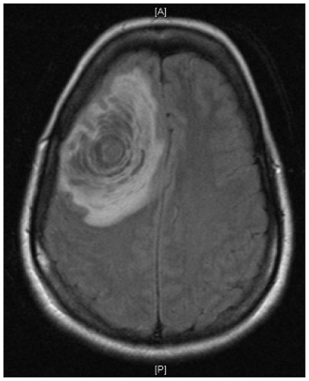
A T1-weighted magnetic resonance imaging scan showed a right frontal lesion with midline shift.
References
-
- Given CA, 2nd, Stevens BS, Lee C. The MRI appearance of tumefactive demyelinating lesions. AJR Am J Roentgenol. 2004;182:195–9. - PubMed
-
- Tan HM, Chan LL, Chuah KL, et al. Monophasic, solitary tumefactive demyelinating lesion: neuroimaging features and neuropathological diagnosis. Br J Radiol. 2004;77:153–6. - PubMed
-
- Mao-Draayer Y, Braff S, Pendlebury W, et al. Treatment of steroid-unresponsive tumefactive demyelinating disease with plasma exchange. Neurology. 2002;59:1074–7. - PubMed
Publication types
MeSH terms
LinkOut - more resources
Full Text Sources