Review
. 2010 Jul;22(7):1003-12.
doi: 10.1016/j.cellsig.2010.01.013. Epub 2010 Jan 25.

Examination of the expanding pathways for the regulation of p21 expression and activity

Affiliations
Review

Examination of the expanding pathways for the regulation of p21 expression and activity

Yong-Sam Jung et al. Cell Signal. 2010 Jul.

Abstract

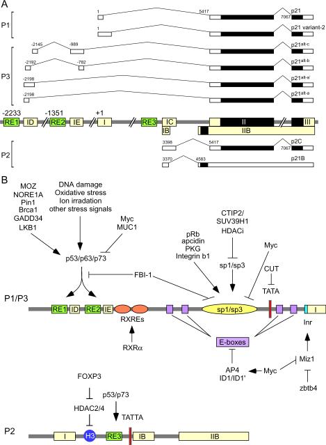
p21(Waf1/Cip1/Sdi1) was originally identified as an inhibitor of cyclin-dependent kinases, a mediator of p53 in growth suppression and a marker of cellular senescence. p21 is required for proper cell cycle progression and plays a role in cell death, DNA repair, senescence and aging, and induced pluripotent stem cell reprogramming. Although transcriptional regulation is considered to be the initial control point for p21 expression, there is growing evidence that post-transcriptional and post-translational regulations play a critical role in p21 expression and activity. This review will briefly discuss the activity of p21 and focus on current knowledge of the determinants that control p21 transcription, mRNA stability and translation, and protein stability and activity.


Figures

Figure 1
Figure 1
The role of p21 in cell survival and death. Under non-stressed conditions, p21 is expressed at low levels and promotes cell cycle progression. Under stress conditions, p21 expression is increased through p53-dependent and -independent pathways. Increased p21 interacts with and inhibits cyclin-CDKs activity. In most tumor cells, the cell cycle checkpoint function of p21 is lost due to p53 mutations. Akt1 phosphorylates and then stabilizes p21 protein for cell survival. P21 inhibits apoptosis by interacting with proapoptotic molecules such as caspase-3 and ASK1.
Figure 2
Figure 2
Human p21 locus. (A) Schematic presentation of various known p21 transcripts. The position of the first nucleotide of p21 transcript (GenBank Accession # Z85996) in the genomic DNA is assigned as the start site of the p21 locus. p21 is the classical p21Waf1/Cip1/Sdi1 transcript [5]. The alternate transcripts, p21variant-2, p21alt-a, p21alt-b, p21alt-c, and p21alt-a1, are presented as reported previously [95]. p21B and p21C are also presented as reported previously [93]. The protein coding regions are marked with solid black boxes. (B) Schematics of the p21 promoters along with various responsive elements, transcription factors, and upstream signals. Please see text for details.
Figure 3
Figure 3
Regulation of p21 by various microRNAs (miRNAs). (A) Schematic diagram showing the genomic structure of five miRNA clusters. miR-106a and miR-17 clusters are a paralogue of miR-106b cluster. miR-19-92 cluster is related to miR-106a-92 cluster. P53 is also differentially regulated by miR-125 and miR-29 clusters. (B) Multiple sequence alignment of six miRNAs. All of these miRNAs share a core seed region, nt 2–8.
Figure 4
Figure 4
Regulation of p21 by RNA-binding proteins (RBPs). Several groups of RBPs are found to regulate p21 expression. Alternative names are included in parentheses. These RBPs contain one or more RNA-binding motifs, including RNA recognition domain (RRM), hnRNP K homology domain (KH), arginine/glycine-rich box (RGG), and double stranded RNA binding motif (dsRBD).
Figure 5
Figure 5
Phosphorylation sites in p21 protein. P21 has six well-defined functional domains, including two cyclin-binding domains (Cy1 and Cy2), a kinase-binding domain (CDK), a Helix motif (Helix), a PCNA-binding domain (PCNA), and a nuclear localization signal (NLS). P21 polypeptide contains 20 serine/threonine residues, seven of which are found to be phosphorylated by various protein kinases indicated at the top of the figure. Five other serine and threonine residues are potential phosphorylation sites (S31, T97, S114, S123, and S137) as predicted by NetPhos 2.0 program (Technical University of Denmark).

References

    1. Sherr CJ. Science. 1996;274(5293):1672–1677. - PubMed
    1. Sherr CJ, Roberts JM. Genes Dev. 1995;9(10):1149–1163. - PubMed
    1. Sherr CJ, Roberts JM. Genes Dev. 1999;13(12):1501–1512. - PubMed
    1. Harper JW, Adami GR, Wei N, Keyomarsi K, Elledge SJ. Cell. 1993;75(4):805–816. - PubMed
    1. el-Deiry WS, Tokino T, Velculescu VE, Levy DB, Parsons R, Trent JM, Lin D, Mercer WE, Kinzler KW, Vogelstein B. Cell. 1993;75(4):817–825. - PubMed

Publication types

MeSH terms

Substances