Review
. 2010 May;29(3):208-48.
doi: 10.1016/j.preteyeres.2010.01.002. Epub 2010 Jan 25.

Novel aspects of corneal angiogenic and lymphangiogenic privilege

Affiliations
Review

Novel aspects of corneal angiogenic and lymphangiogenic privilege

David Ellenberg et al. Prog Retin Eye Res. 2010 May.

Abstract

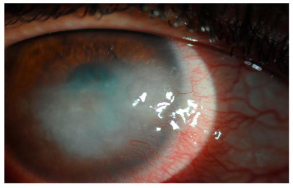
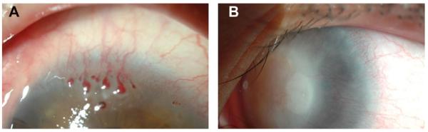
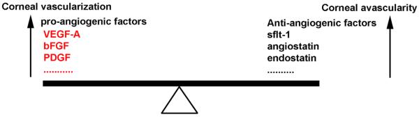
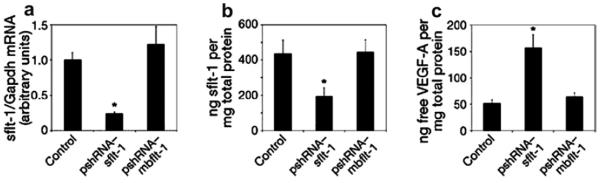
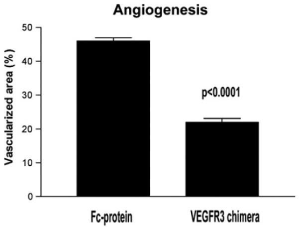
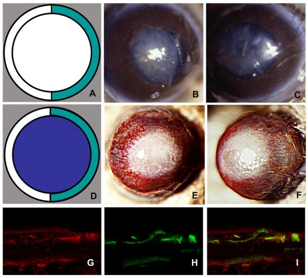
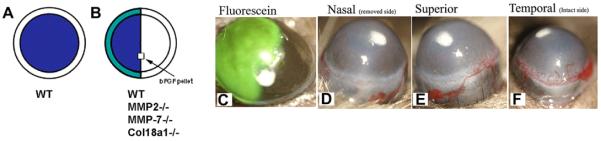
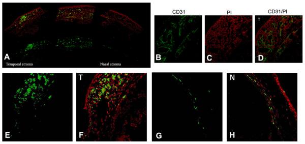
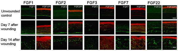
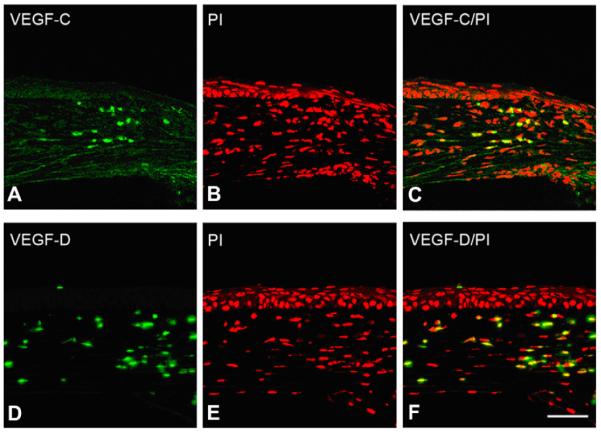
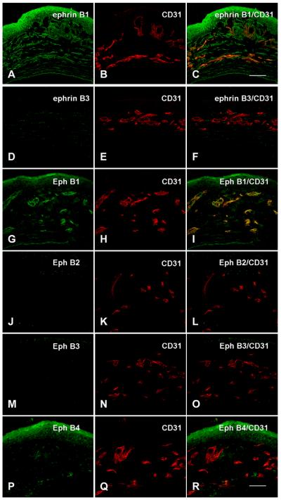
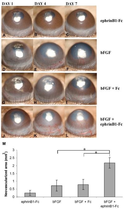
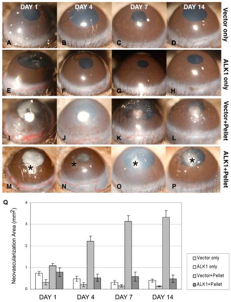
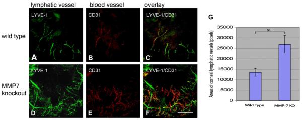
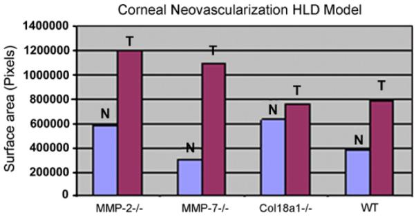
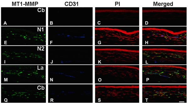
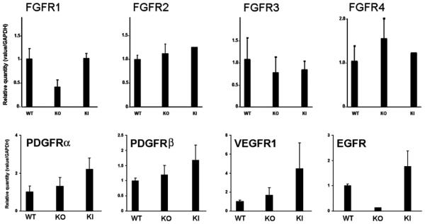
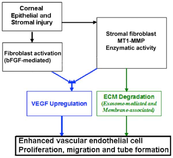
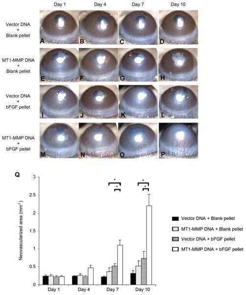
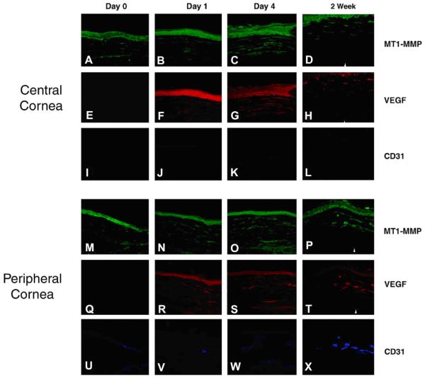
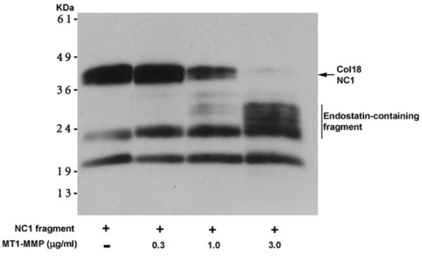
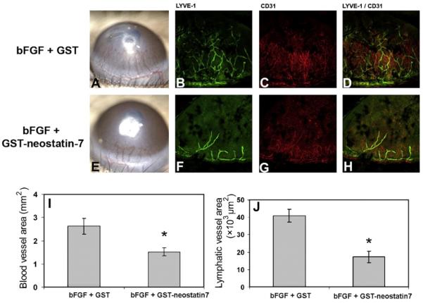
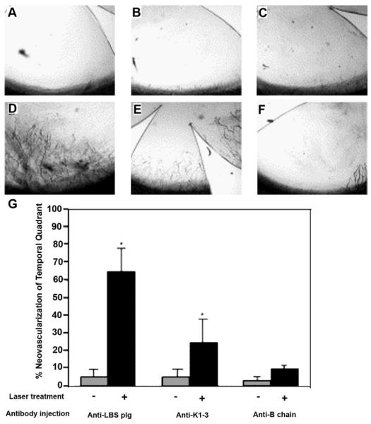
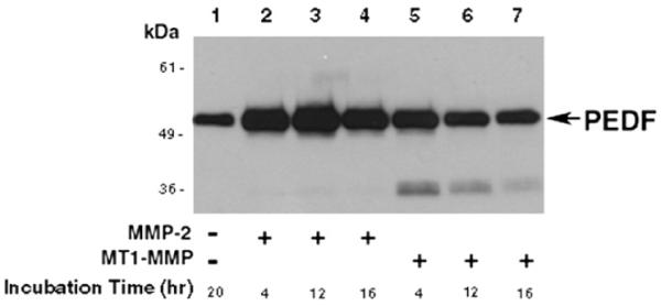
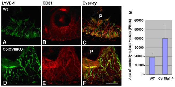
In this article, we provide the results of experimental studies demonstrating that corneal avascularity is an active process involving the production of anti-angiogenic factors, which counterbalance the pro-angiogenic/lymphangiogenic factors that are upregulated during wound healing. We also summarize pertinent published reports regarding corneal neovascularization (NV), corneal lymphangiogenesis and corneal angiogenic/lymphangiogenic privilege. We outline the clinical causes of corneal NV, and discuss the angiogenic proteins (VEGF and bFGF) and angiogenesis regulatory proteins. We also describe the role of matrix metalloproteinases MMP-2, -7, and MT1-MMP, anti-angiogenic factors, and lymphangiogenic regulatory proteins during corneal wound healing. Established and potential new therapies for the treatment of corneal neovascularization are also discussed.


Figures

Fig. 1
Fig. 1
Clinical appearance of corneal NV in inflammatory disorders. A: Corneal NV in Salzmann's nodular degeneration. B: Corneal NV due to Rosacea. Dilated vessels at the limbus advance into the cornea predominantly inferiorly.
Fig. 2
Fig. 2
Clinical appearance of corneal NV post-keratoplasty (A) and its magnification (B).
Fig. 3
Fig. 3
Clinical appearance of corneal NV in corneal infections. A: Superficial corneal vessel growth associated with Acanthamoeba Keratitis infection. The vast majority of reported cases of Acanthamoeba keratitis have been associated with contact lens use. Vessels normally grow inferiorly into the central cornea. This amebic infection has rarely been seen to spread beyond the cornea to affect the perilimbal and posterior ocular structures. However, a recent report shows that some patients had unexpected histopathologic findings of diffuse neuroretinal ischemia and perivascular lymphocytic infiltrates, some with vascular thrombosis, and chronic chorioretinal inflammation (Awwad et al., 2007). B: Retro-illumination image of an eye with deep stromal corneal NV due to interstitial keratitis. Patients with interstitial keratitis experience pain, photophobia, increased tearing, blepharospasm, and decreased vision. Neovascularization progresses centrally over time until the vessels coalesce in the central cornea.
Fig. 4
Fig. 4
Corneal NV in contact lens-related hypoxia.
Fig. 5
Fig. 5
Superficial and mid stromal corneal NV due to neurotrophic ulceration.
Fig. 6
Fig. 6
Corneal inflammation and neovascularization associated with limbal stem cell deficiency (A and B). Corneal NV associated with this condition is clinically challenging in that it persists long after the insult, and may not improve without transplantation of limbal stem cells.
Fig. 7
Fig. 7
A diagram depicting corneal angiogenesis and avascularity. The normally quiescent vasculature (corneal avascularity) can be activated to sprout new capillaries (corneal vascularization), a process controlled by an angiogenic switch mechanism. In some pathologies not causing corneal angiogenesis, the absence of angiogenic inducers may preserve corneal avascularity, while in others the angiogenic inducers are present but held in check by higher levels of angiogenic inhibitors. An increase in the levels of pro-angiogenic factors in the cornea tilts the balance towards corneal vascularization. An increase in the levels of anti-angiogenic factors in the cornea tilts the balance towards corneal avascularity (Modified from Hanahan and Folkman, 1996).
Fig. 8
Fig. 8
Real-time RT-PCR reveals reduced levels of sflt-1 mRNA (a) and an enzyme-linked immunosorbent assay (ELISA) reveals reduced levels of sflt-1 protein (b) and increased free VEGF-A protein (c) in WT mouse corneas 3 days after injection of pshRNA-sflt-1, but not pshRNA-mbflt-1. The asterisk denotes P < 0.05, Bonferroni corrected Mann–Whitney U-test. n = 8–12. Error bars depict s.e.m. (reprinted with permission from Ambati et al., 2006).
Fig. 9
Fig. 9
Diminished corneal vessels after administration of a VEGFR-3 chimera to wounded corneas. The neovascular response after cautery of de-epithelialized corneas is significantly diminished when a VEGFR-3 chimeric protein is administered locally (reprinted with permission from Cursiefen et al., 2006a).
Fig. 10
Fig. 10
Hemilimbal deficiency: a model for injury-induced corneal NV. The diagrams depict limbal injury (A; green) and limbal plus epithelial removal (D; purple). The nasal limbi of WT mouse corneas were removed and photographed at day 7 after surgery (B, C). The nasal limbus and the epithelium of WT mouse corneas were removed and the corneas photographed at day 7 after surgery (E, F). Vascularized vessels were immunostained with anti-type IV collagen (G), anti-CD31 antibodies (H), and double staining (I) (reprinted with permission Azar, 2006).
Fig. 11
Fig. 11
A diagram of the hemilimbal barrier model. Removal of the corneal epithelial layer by #15 blade (A) and hemilimbal (B; half of the limbal and corneal epithelium with bFGF-pellet implantation). Corneas of hemilimbal wounding were stained with fluorescein (C). Corneal NV at day 5 post-intrastromal bFGF-pellet implantation and hemilimbal debridement (n = 5). The debrided nasal side has no corneal NV (D), corneal NV were developed from the temporal unwounded side (F).
Fig. 12
Fig. 12
Enhanced corneal CD31 and VEGF-A expression in hemilimbal wounded corneas. Wounded corneas were immunostained with anti-CD31 (B) and VEGF A (E; temporal side, G; nasal side) antibodies. Enhanced CD31 immunostaining in the temporal side of the unwounded corneas (B), PI staining (C), and merged images (D). Enhanced corneal VEGF-A expression in the temporal side of the unwounded cornea (E) compared to the wounded side (nasal side) of the cornea (bar = 50 μm).
Fig. 13
Fig. 13
Enhanced expression of FGF1, 2, 3, 7, and 22 was detected in alkali wounded corneas. Corneas were treated with 1 N NaOH for 1 min and harvested at days 7 and 14 (n = 5). Corneal sections were immunostained with anti-FGF, 1, 2, 3, 7, and 22 antibodies (bar = 50 μm).
Fig. 14
Fig. 14
bFGF induces corneal VEGF-C/-D expression. Mouse corneas were implanted with bFGF pellets and corneal sections were immunostained with anti-VEGF-C and -D antibodies. VEGF-C/-D expression was shown to be enhanced after bFGF-pellet implantation (bar = 50 μm).
Fig. 15
Fig. 15
Expression of Eph and ephrin in bFGF-induced corneas. Corneas were harvested at day 7 after bFGF-pellet implantation (n = 3). Corneal sections were double immunostained with anti-ephrinB1, B3, EphB1, B2, B3, B4, and with CD31 antibodies. Notably, EphB1 is co-localized with CD31 in the corneal vessels after bFGF implantation (bar = 50 μm).
Fig. 16
Fig. 16
Effect of ephrinB1-Fc on corneal pocket assay. The pellet containing ephrinB1-Fc (A–C), bFGF (50 ng/pellet) (D–), bFGF + Fc (G–I), and ephrinB1-Fc + bFGF (J–L) was inserted into corneal stromal pocket. Photographs were taken on days 1, 4, and 7 after implantation. bFGF-induced corneal NV was significantly enhanced by ephrinB1-Fc, in vivo. M: Seven days after pellet insertion, the area of corneal NV was calculated using NIH ImageJ software. Results are representative of at least three independent experiments and represent the mean ± SEM. *P < 0.05 (reprinted with permission from Kojima et al., 2007a).
Fig. 17
Fig. 17
bFGF-induced corneal NV is inhibited by naked ALK-1 DNA injection, in vivo. No-pellet controls are shown in A–H: Injection of naked DNA [ALK-1 (E–H) and vector only (A–D)] did not induce corneal NV. The vector plus pellet positive controls are shown in I–L: Development of NV in the corneal stroma was evident by day 4 (J); new vessels continued to grow in the direction of the pellet on days 7 and 14. None of the mice in the ALK-1 and pellet groups (M–P) showed development of corneal NV on days 1, 4, 7, and 14 after pellet implantation. Asterisk (*) indicates pellet implantation. The area of corneal NV of the four groups at days 1–14 is shown (Q) (reprinted with permission from Albe et al., 2005).
Fig. 18
Fig. 18
Differential HVS protein expression in postnatal days 1 and 16. Representative 2-DE gels of proteins obtained from the lens and vitreous of P1 mouse and P16 mouse using IPG strips with pH range 4–7 and 7–10. The proteins excised for analysis and identification by MS are marked with numbers from 1 to 46 (reprinted with permission from Albe et al., 2008).
Fig. 19
Fig. 19
Enhanced bFGF-induced corneal angiogenesis and lymphangiogenesis in MMP-7−/− mice. A bFGF pellet was implanted into WT and MMP-7−/− mice for 7 days. Three mouse corneas were whole-mounted and double immunostained with anti-LYVE (A, D) and CD31 (B, E) antibodies. The areas of corneal angiogenic and lymphangiogenic vessels were determined using ImageJ. The levels of corneal lymphangiogenesis in MMP-7−/− are significantly higher than WT mice.
Fig. 20
Fig. 20
The total surface area of corneal NV in MMP-2−/− , MMP-7−/−, Col18a1−/− (collagen XVIII), or WT (wild type) mice in the hemilimbal deficiency model. At day 7 after injury, total areas of corneal NV in temporal side (T) and nasal side (N) of the wounded corneas. (HLD: Hemilimbal Deficiency).
Fig. 21
Fig. 21
Enhanced corneal MT1-MMP and CD31 expression after alkali wounding. A mouse was anesthetized by intraperitoneal injection of ketamine HC1 (50 mg/kg) and xylazine (10 mg/kg). Topical tetracaine was applied to the cornea for 1 min before alkali wounding. A Whatman #3 filter cut by a trephine of 2-mm diameter was presoaked with 2 μL of 1 N sodium hydroxide solution, and placed on the central cornea of left eye for 1 min. Wounded surfaces were immediately washed by physiologic saline. Day 7 alkali wounded corneas were immunostained with four different anti-MT1-MMP (N1, N2, La, Cb), CD31, and PI. Enhanced MT1-MMP (E, I, M and Q) and CD31 (F, J, N and R) immunostaining were detected in alkali wounded corneas when compared to unwounded control (A and B) (bar = 50 μm).
Fig. 22
Fig. 22
Diminished FGFR-1 and EGFR in MT1-MMP knockout keratocyte cell lines. Total RNA was extracted from WT, KO, and KI corneal keratocyte cells and quantitative real-time PCR was performed. Diminished FGFR-1 and EGFR expression were detected in MT1-MMP knockout keratocytes and recovered in MT1-MMP knockin keratocytes (Onguchi et al., 2009).
Fig. 23
Fig. 23
The diagram depicts MT1-MMP functions during corneal wounding. Corneal kertocyte MT1-MMP upregulates VEGF production, MMP-2 activation, and cleavage of ECM proteins after bFGF-pellet implantation and alkali wounding.
Fig. 24
Fig. 24
Enhanced bFGF-induced corneal NV after a combination of bFGF-pellet implantation and naked MT1-MMP DNA plasmid injection. The blank pellets were implanted immediately after MT1-MMP DNA (E–H) or vector control DNA (A–D) was injected into corneal stroma. Likewise, the bFGF pellets were implanted immediately after MT1-MMP DNA (M–P) or vector control DNA (I–L) was injected into corneal stroma. Photographs were taken on days 1, 4, 7, and 10 after surgery. Q: Graphic representation of at least five independent experiments (mean ± SEM, *P < 0.05) (reprinted with permission from Onguchi et al., 2009).
Fig. 25
Fig. 25
Enhanced MT1-MMP expression in central and peripheral corneas at day 4 after bFGF implantation. bFGF-induced vessels were detected at peripheral cornea, but not in the central cornea at day 4 and 14. Enhanced VEGF expression was detected at day 4 and 14 after bFGF-pellet implantation (reprinted with modification from Azar, 2006).
Fig. 26
Fig. 26
MT1-MMP cleaves a recombinant collagen NC1 fragment. MT1-MMP cleaves a recombinant NC1 (Chang et al., 2005) fragment of collagen XVIII to generate endostatin-containing fragments. In an in vitro assay, the addition of higher concentrations of MT1-MMP enhanced the production of endostatin-containing fragments detected by anti-endostatin antibody (Chang et al., 2005).
Fig. 27
Fig. 27
GST-neostatin-7 reduced the bFGF-induced corneal hem- and lymphangiogenesis. Mouse corneas were implanted with bFGF (80 ng/pellet) plus either GST (500 ng/pellet) (A) or GST-neostatin-7 protein (500 ng/pellet) (B). Corneal NV images were taken by slit-lamp microscope 7 days after pellet implantation. The pellet containing GST-neostatin-7 significantly reduced bFGF-induced NV (I). Corneal lymphangiogenesis was visualized by whole-mount immunohistochemical staining using anti-LYVE-1 antibody on day 7 after pellet implantation. Enhanced corneal lymphangiogenesis was visualized in GST plus bFGF implanted corneas (B); however, diminished corneal lymphatic vessels were observed in GST-neostatin-7 plus bFGF implanted corneas (F). Overlay images of corneal angiogenesis and lymphangiogenesis are shown in (D) and (H), respectively. Quantification of lymphatic vessels showed a reduction in corneal lymphatic vessels in GST-neostatin-7 implanted corneas (J). *Represents P < 0.05 (reprinted with permission from Kojima et al., 2008).
Fig. 28
Fig. 28
Corneal injection of anti-angiostatin antibody enhanced corneal vascularization after excimer laser wounding. Intrastromal injection of anti-plasminogen (LBS), anti-angiostatin, and anti-plasmin B chain antibodies in unwounded (A–C) and wounded (D–E) rat corneas. Corneal NV was visualized after India ink perfusion (flat mounted corneas). Quotidian injection (for 5 days) of anti-plasminogen (LBS; A, D) and anti K-1–3 (B, E) antibodies induced corneal NV after wounding (D, E), but not in unwounded corneas (A, B). In contrast, the injection of an anti-plasmin B chain showed no significant increase of vascularization in unwounded (C) and wounded (F) corneas. (G) Quantification of corneal NV and statistical analysis (unpaired t-test). (*, statistically significant) (reprinted with permission from Gabison et al., 2004).
Fig. 29
Fig. 29
In vitro degradation of PEDF by MT1-MMP. Recombinant PEDF was incubated with active MMP-2 or MT1-MMP. The degradation products of PEDF by MT1-MMP were detected by western blot analysis using anti-PEDF antibodies.
Fig. 30
Fig. 30
VEGF-D enhances corneal lymphangiogenesis in col18a1−/− mice. Corneas from WT and col18a1−/− mouse were implanted with VEGF-D pellets (160 ng/pellet). Corneas were harvested 7 days later and processed for whole-mount immunostaining. LYVE-1 (A and D) and CD31 immunostaining (B and E) are shown. Quantification of LYVE-1 staining revealed that more lymphatic vessels developed in the pellet-implanted side of col18a1 knockout mouse corneas than in the pellet-implanted side of WT corneas (G) (bar = 50 μm).

Similar articles

Cited by

References

    1. Abbas A, Khan B, Feroze AH, Hyman GF. Thalidomide prevents donor corneal graft neovascularization in an alkali burn model of corneal angiogenesis. J. Pak. Med. Assoc. 2002;52:476–482. - PubMed
    1. Abdiu O, Van Setten G. Antiangiogenic activity in tears: presence of pigment-epithelium-derived factor. New insights and preliminary results. Ophthalmic Res. 2008;40:16–18. - PubMed
    1. Abedi H, Zachary I. Vascular endothelial growth factor stimulates tyrosine phosphorylation and recruitment to new focal adhesions of focal adhesion kinase and paxillin in endothelial cells. J. Biol. Chem. 1997;272:15442–15451. - PubMed
    1. Acuff HB, Sinnamon M, Fingleton B, Boone B, Levy SE, Chen X, Pozzi A, Carbone DP, Schwartz DR, Moin K, Sloane BF, Matrisian LM. Analysis of host- and tumor-derived proteinases using a custom dual species microarray reveals a protective role for stromal matrix metalloproteinase-12 in non-small cell lung cancer. Cancer Res. 2006;66:7968–7975. - PubMed
    1. Adamis AP, Meklir B, Joyce NC. In situ injury-induced release of basic-fibroblast growth factor from corneal epithelial cells. Am. J. Pathol. 1991;139:961–967. - PMC - PubMed

Publication types

MeSH terms

Substances