. 2010 May;38(9):2931-43.
doi: 10.1093/nar/gkq011. Epub 2010 Jan 25.

The novel mouse Polo-like kinase 5 responds to DNA damage and localizes in the nucleolus

Affiliations

The novel mouse Polo-like kinase 5 responds to DNA damage and localizes in the nucleolus

Zdenek Andrysik et al. Nucleic Acids Res. 2010 May.

Abstract

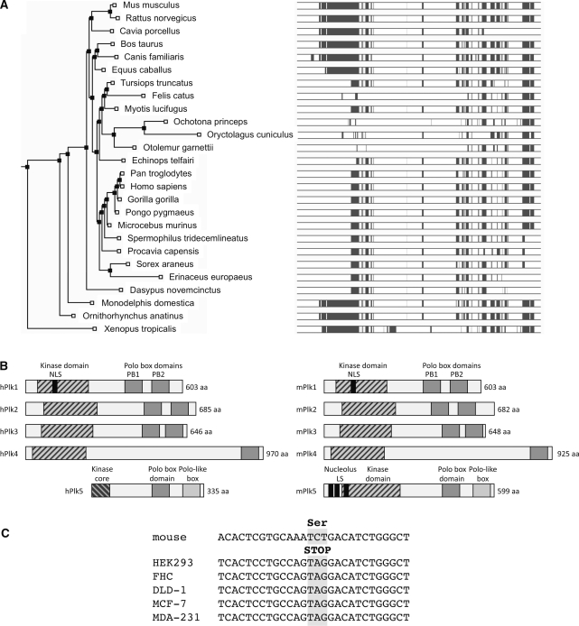
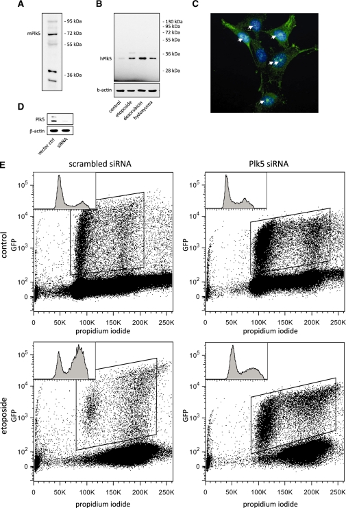
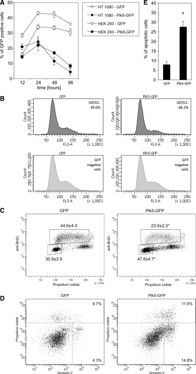
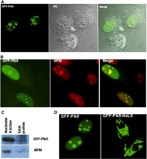
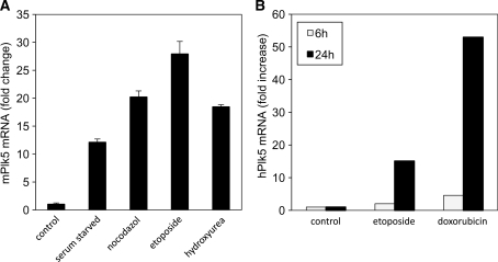
Polo-like kinases (Plk1-4) are emerging as an important class of proteins involved in many aspects of cell cycle regulation and response to DNA damage. Here, we report the cloning of a fifth member of the polo-like kinase family named Plk5. DNA and protein sequence analyses show that Plk5 shares more similarities with Plk2 and Plk3 than with Plk1 and Plk4. Consistent with this observation, we show that mouse Plk5 is a DNA damage inducible gene. Mouse Plk5 protein localizes predominantly to the nucleolus, and deletion of a putative nucleolus localization signal (NoLS) within its N-terminal moiety disrupts its nucleolar localization. Ectopic expression of Plk5 leads to cell cycle arrest in G1, decreased DNA synthesis, and to apoptosis, a characteristic it shares with Plk3. Interestingly, in contrast to mouse Plk5 gene, the sequence of human Plk5 contains a stop codon that produces a truncated protein lacking part of the kinase domain.


Figures

Figure 1.
Figure 1.
Structural organization of mouse and human Plk5. (A) A rooted tree was constructed using PHYLIP (70) showing one model of evolutionary descent. (B) Shared domains between mPlk5, hPlk5 and the other Plks. NoLS indicates the nucleolus localization signal. (C) Plk5 DNA sequence from different human cell lines of cancer and noncancer origin showing the presence of the nonsense codon in exon 6. The cell lines include: HEK-293 (Human embryonic kidney cell line), FHC (normal human fetal colon cell line), DLD-1 (human colon carcinoma), MCF-7 (breast cancer cell line) and MDA-231 (breast cancer cell line). The mouse Plk5 sequence is also shown for comparison.
Figure 2.
Figure 2.
Human and mouse Plk5 proteins are both expressed in cultured cells. Western blot showing that mouse Plk5 (A) and human Plk5 (B) proteins are detected in lysates from NIH3T3 and HEK-293 cells, respectively, using a Plk5 polyclonal antibody. (C) Immunofluorescence detection of endogenous mPlk5 in discrete foci (arrows) in NIH3T3 cells. (D) A shRNA that targets hPlk5 mRNA was able to deplete Plk5 protein from HEK-293 cells. (E) HEK-293 cells transiently transfected with vector expressing both shRNA against Plk5 and GFP were analyzed by flow cytometry. The cell cycle distribution of GFP-positive cells is shown in left top corner of each panel. Depletion of hPlk5 from HEK-293cells leads to loss of G2/M checkpoint in response to treatment with the DNA damaging agent, etoposide (lower panel, right) compared to nondepleted cells (lower panel, left).
Figure 3.
Figure 3.
Ectopic expression of murine Plk5 induces G1 arrest and cell death by apoptosis. (A) Growth curves of cells transfected by either a pEGFP plasmid control or a pEGFP-mPlk5 construct. (B) Cell cycle profile of cells transfected with either pEGFP or pEGFP-mPlk5 showing G1 arrest in the Plk5 transfected cells. (C) BrdU staining of cells transfected by either pEGFP or pEGFP-mPlk5 confirms the G1 arrest in the Plk5 transfected cells (upper panel, right) compared to GFP transfected cells (upper panel, left). No major change is seen in GFP negative cells (lower panel). (D) Annexin staining of the transfected cells showing increased apoptosis in pEGFP-mPlk5 transfected cells and (E) Quantitave analysis of nuclei with apoptotic morphology as a result of mPlk5 expression confirming the Annexin V staining. Asterisk denotes statistical difference P < 0.05.
Figure 4.
Figure 4.
mPlk5 localizes to the nucleolus. (A) GFP-mPlk5 localizes to discrete foci. (B) GFP-mPlk5 co-localization with endogenous NPM in the nucleolus. (C) Enrichment for mPlk5 in the nucleolar fraction. (D) Loss of nucleolar localization in the NoLS mutant.
Figure 5.
Figure 5.
Plk5 expression is induced following different stress stimuli. (A) Murine NIH 3T3 cells were either left untreated (control) or treated for 18 h with the DNA damaging agents etoposide (Etop) or HU, the spindle disassembly agent, nocodazole (Noc) or were serum starved (SS). (B) Human HEK293 cells were either left untreated (control) or treated with DNA damaging agent etoposide or doxorubicin for 6 or 24 h. The Plk5 mRNA level was measured by qPCR in both cases.
Figure 6.
Figure 6.
Plk5 transcriptional activation in response to DNA damage is not p53-dependent. (A) p53 consensus binding sites in Plk5 promoter, (B) mPlk5 mRNA expression levels measured by qPCR following etoposide and pifithrin treatment of NIH 3T3 cells for 18 h. (C) Expression of p21 protein in lysates from cells either nontreated (1), treated with pifithrin (2), etoposide alone (3) or pifithrin and etoposide (4) was analyzed by western blot.

Similar articles

Cited by

References

    1. Lee KS, Grenfell TZ, Yarm FR, Erikson RL. Mutation of the polo-box disrupts localization and mitotic functions of the mammalian polo kinase Plk. Proc. Natl Acad. Sci. USA. 1998;95:9301–9306. - PMC - PubMed
    1. Logarinho E, Sunkel CE. The Drosophila POLO kinase localises to multiple compartments of the mitotic apparatus and is required for the phosphorylation of MPM2 reactive epitopes. J. Cell Sci. 1998;111(Pt 19):2897–2909. - PubMed
    1. Shirayama M, Zachariae W, Ciosk R, Nasmyth K. The Polo-like kinase Cdc5p and the WD-repeat protein Cdc20p/fizzy are regulators and substrates of the anaphase promoting complex in Saccharomyces cerevisiae. EMBO J. 1998;17:1336–1349. - PMC - PubMed
    1. Qian YW, Erikson E, Li C, Maller JL. Activated polo-like kinase Plx1 is required at multiple points during mitosis in Xenopus laevis. Mol. Cell Biol. 1998;18:4262–4271. - PMC - PubMed
    1. Moutinho-Santos T, Sampaio P, Amorim I, Costa M, Sunkel CE. In vivo localisation of the mitotic POLO kinase shows a highly dynamic association with the mitotic apparatus during early embryogenesis in Drosophila. Biol. Cell. 1999;91:585–596. - PubMed

Publication types

Substances