. 2010 Mar 19;285(12):8985-94.
doi: 10.1074/jbc.M109.078725. Epub 2010 Jan 25.

Outer membrane protein I of Pseudomonas aeruginosa is a target of cationic antimicrobial peptide/protein

Affiliations

Outer membrane protein I of Pseudomonas aeruginosa is a target of cationic antimicrobial peptide/protein

Yu-Min Lin et al. J Biol Chem. .

Abstract

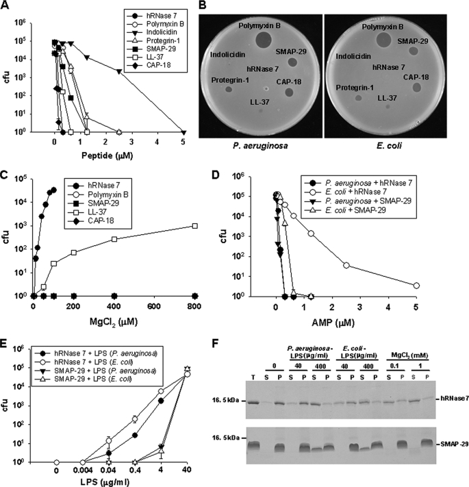
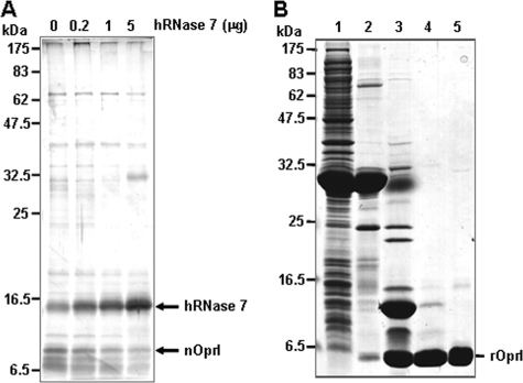
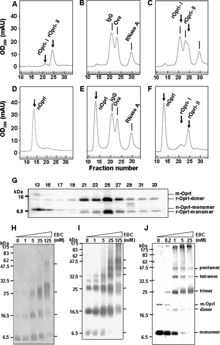
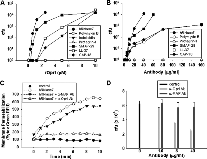
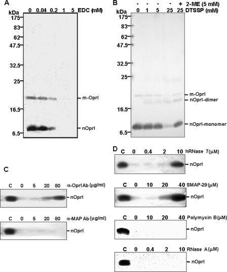
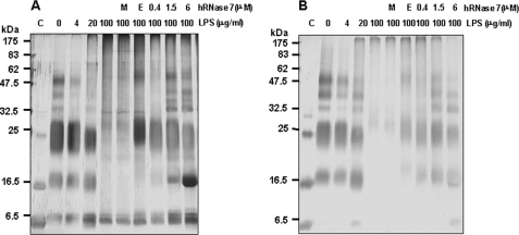
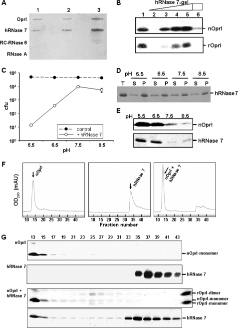
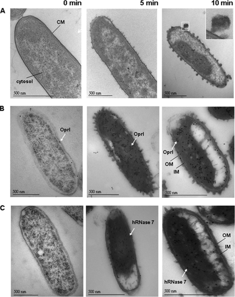
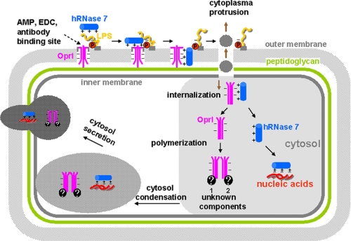
Cationic antimicrobial peptides/proteins (AMPs) are important components of the host innate defense mechanisms against invading microorganisms. Here we demonstrate that OprI (outer membrane protein I) of Pseudomonas aeruginosa is responsible for its susceptibility to human ribonuclease 7 (hRNase 7) and alpha-helical cationic AMPs, instead of surface lipopolysaccharide, which is the initial binding site of cationic AMPs. The antimicrobial activities of hRNase 7 and alpha-helical cationic AMPs against P. aeruginosa were inhibited by the addition of exogenous OprI or anti-OprI antibody. On modification and internalization of OprI by hRNase 7 into cytosol, the bacterial membrane became permeable to metabolites. The lipoprotein was predicted to consist of an extended loop at the N terminus for hRNase 7/lipopolysaccharide binding, a trimeric alpha-helix, and a lysine residue at the C terminus for cell wall anchoring. Our findings highlight a novel mechanism of antimicrobial activity and document a previously unexplored target of alpha-helical cationic AMPs, which may be used for screening drugs to treat antibiotic-resistant bacterial infection.


Figures

FIGURE 1.
FIGURE 1.
Effect of LPS on the bactericidal activity of AMPs. A, effect of AMPs on viability of P. aeruginosa cells. Bacteria (5–10 × 104 cfu) were treated with AMP at 37 °C for 3 h and then plated for the determination of the remaining cfu (17). B, the susceptibility of P. aeruginosa to various AMPs (4 μg each dotted on the plate) was assayed by the formation of an inhibition zone on Luria-Bertani agar plates. C, effect of MgCl2 on the bactericidal activity of AMPs to P. aeruginosa cells. D, viability of P. aeruginosa and E. coli cells with hRNase 7 and SMAP-29 treatment. E, effect of hRNase 7 and SMAP-29 on viability of P. aeruginosa in the presence of different LPS. F, effect of MgCl2 and LPS on the binding of hRNase 7 and SMAP-29 to P. aeruginosa. hRNase 7 (40 μg/ml, top panel), and SMAP-29 (160 μg/ml, bottom panel) were used for the binding assays in the presence of LPS and MgCl2 as indicated. T, S, and P represent the total protein, supernatant, and pellet, respectively, after centrifugation.
FIGURE 2.
FIGURE 2.
Identification and production of OprI. A, identification of hRNase 7-binding proteins. The membrane extracts of P. aeruginosa (2 μg) were incubated with 10 μl of hRNase 7 gel in the presence of increasing amounts of free hRNase 7 and subjected to SDS-PAGE and silver staining. B, analysis of rOprI by 14% SDS-PAGE and Coomassie Blue staining. Lane 1, crude lysate from E. coli BL21(DE3) transformed with thioredoxin/OprI-fused gene; lane 2, eluate of nickel affinity column; lane 3, digestion product of protease Factor Xa; lane 4, flowthrough of second nickel affinity column; lane 5, eluate of fast protein liquid chromatography SuperoseTM 12 column gel filtration chromatography.
FIGURE 3.
FIGURE 3.
Polymeric structure of OprI. Gel filtration chromatographic analyses of rOprI (A), molecular size marker proteins (B), and both (C). Purified rOprI as well as immunoglobulin (150 kDa), ovalbumin (44 kDa), and bovine RNase A (14 kDa), 100 μg each, were employed for the analyses. D–F, gel filtration chromatographic analysis of nOprI (D), nOprI and molecular size marker proteins (E), and nOprI and rOprI (F). G, the nOprI and rOprI in the column eluates of F were analyzed by 14% SDS-PAGE and Western blotting. H and I, rOprI polymer levels (0.75 μg) cross-linked with increasing amounts of EDC (as indicated) at pH 5.5 for 10 min underwent 14% SDS-PAGE and silver staining (H) and then Western blot analysis (I). J, nOprI polymer levels (4 μg) cross-linked with EDC underwent Western blot analysis. m-OprI, modified native OprI.
FIGURE 4.
FIGURE 4.
Role of OprI in membrane permeability and susceptibility to α-helical AMPs. A, effect of rOprI on the viability of P. aeruginosa cells (1 × 10⁵ cfu) treated with 0.6 μm hRNase 7, 1.2 μm polymyxin B, 5 μm indolicidin, 2.5 μm protegrin-1, 1.2 μm SMAP-29, 0.84 μm LL-37, or 0.25 μm CAP-18. The values of polymyxin B, indolicidin, and protegrin-1 are superimposable at 100 cfu. B, protection of P. aeruginosa from bactericidal activities of AMPs on pretreatment with anti-OprI antibody. C, inhibition of 2.5 μm hRNase 7-induced membrane permeability by anti-OprI and anti-MAP antibody (80 μg/ml). D, inhibition of P. aeruginosa growth by anti-OprI antibody. RFU, relative fluorescence units of SYTOX® Green.
FIGURE 5.
FIGURE 5.
Association of OprI with surface components. A and B, analysis of OprI complex after cross-linking. Small aliquots of P. aeruginosa were suspended in 10 mm sodium phosphate, pH 5.5 and 7.5, respectively, with increasing concentrations of EDC (A) or DTSSP (B). The bacteria underwent nonreducing SDS-PAGE and then Western blot analysis except for the one marked with 2-mercaptoethanol (2-ME). C and D, inhibition of EDC cross-linking. The bacteria were incubated with antibodies (anti-OprI and anti-MAP, C) or AMPs (hRNase 7, SMAP-29, polymyxin B, and bovine RNase A, D) before the addition of 5 mm EDC. m-OprI, modified native OprI.
FIGURE 6.
FIGURE 6.
Dissociation of OprI-LPS complex by hRNase 7 in vitro. rOprI (2 μg) incubated with increasing amounts of LPS for 10 min was cross-linked with 125 mm EDC for 30 min at pH5.5 and underwent SDS-PAGE and silver staining (A) and then Western blot analysis with anti-OprI antibody (B). The OprI-LPS (100 μg/ml) mixture was treated with 1 mm MgCl2 (M), 1 mm EDTA (E), and various amounts of hRNase 7 before cross-linking and further analyses. C represents rOprI without EDC treatment.
FIGURE 7.
FIGURE 7.
Actions of hRNase 7 on OprI. A, recognition of rOprI by ribonucleases. Increasing amounts of rOprI (0.005, 0.05, and 0.5 μg), hRNase 7, bullfrog RC-RNase 6, and bovine RNase A (0.025, 0.25, and 2.5 μg) were blotted on nitrocellulose membrane, incubated with 20 μg of rOprI, and probed with anti-OprI antibody. B, pull-down experiments of nOprI and rOprI by hRNase 7 gel. The membrane extracts (4 μg for top panel) and rOprI (2 μg for bottom panel) were incubated with 0, 5, 10, and 20 μl of hRNase 7 gel suspensions, to which were added 20, 10, 5, and 0 μl of mock Sepharose CL4B gel suspensions (lanes 2–5). The OprIs pulled down by gels were analyzed by Western blotting. 0.7 μg of membrane proteins (top panel) and 0.2 μg of rOprI (bottom panel) were used as a positive control (lane 1). 4 μg of anti-OprI antibody was used to abolish the binding of OprI to hRNase 7 gels (lane 6). C, bactericidal activity of hRNase 7 to P. aeruginosa at different pH values. D, binding of hRNase 7 to P. aeruginosa at different pH values. T, S, and P represent total hRNase 7, supernatant, and pellet, respectively, after centrifugation. E, pH-dependent binding of hRNase 7 to OprI. The membrane extracts were incubated with hRNase 7 gel (top panel), and hRNase 7 was incubated with rOprI gel (bottom panel) in 20 mm sodium phosphate, pH 5.5–8.5. The bound proteins were examined on Western blot analysis with anti-OprI and anti-hRNase 7 antibodies, respectively. F, analyses of nOprI, hRNase 7, and nOprI-hRNase 7 complex by gel filtration chromatography at pH 5.5. G, examination of nOprI and hRNase 7 in column eluates of F by Western blot analysis.
FIGURE 8.
FIGURE 8.
Localization of OprI and hRNase 7 in hRNase 7-treated P. aeruginosa. A, morphological changes of P. aeruginosa after hRNase 7 treatment. P. aeruginosa was treated with hRNase 7 at 37 °C for 5 and 10 min and examined on transmission electron microscopy. B and C, immunohistochemistry of OprI and hRNase 7 protein after hRNase 7 treatment. Thin sections of hRNase 7-treated bacteria were incubated with anti-OprI antibody (B) or anti-hRNase 7 antibody (C), respectively. CM, OM, and IM represent bacterial membrane, outer membrane, and inner membrane, respectively. The dark dots indicated with white arrows in B and C represent OprI and hRNase 7, respectively.
FIGURE 9.
FIGURE 9.
Proposed action mechanism of hRNase 7 on P. aeruginosa. The polymeric α-helical OprIs embed in the outer membrane, which contains fatty acids at the N terminus for membrane integration, an EDC-accessible loop for AMP/LPS recognition, and a C-terminal lysine residue for cell-wall anchoring. The associated LPS and fatty acids of OprI are eliminated on hRNase 7 treatment. On subsequent internalization of OprI along with invading hRNase 7, the membrane is permeable to metabolites. The internalized OprI with anionic surface may be directed to cationic cytoplasmic components and hRNase 7 aggregated anionic nucleic acids. Eventually, all of the condensed cytoplasmic components are exported across the permeabilized membrane before cell death.

Similar articles

Cited by

References

    1. Giamarellou H. (2002) J. Antimicrob. Chemother. 49, 229–233 - PubMed
    1. Zasloff M. (2002) Nature 415, 389–395 - PubMed
    1. Brown K. L., Hancock R. E. (2006) Curr. Opin. Immunol. 18, 24–30 - PubMed
    1. Bowdish D. M., Davidson D. J., Hancock R. E. (2005) Curr. Protein Pept. Sci. 6, 35–51 - PubMed
    1. Gläser R., Harder J., Lange H., Bartels J., Christophers E., Schröder J. M. (2005) Nat. Immunol. 6, 57–64 - PubMed

Publication types

LinkOut - more resources