. 2010 Mar 19;285(12):9090-9.
doi: 10.1074/jbc.M109.080457. Epub 2010 Jan 25.

Biochemical and biological characterization of a new oxidized avidin with enhanced tissue binding properties

Affiliations

Biochemical and biological characterization of a new oxidized avidin with enhanced tissue binding properties

Antonio Verdoliva et al. J Biol Chem. .

Abstract

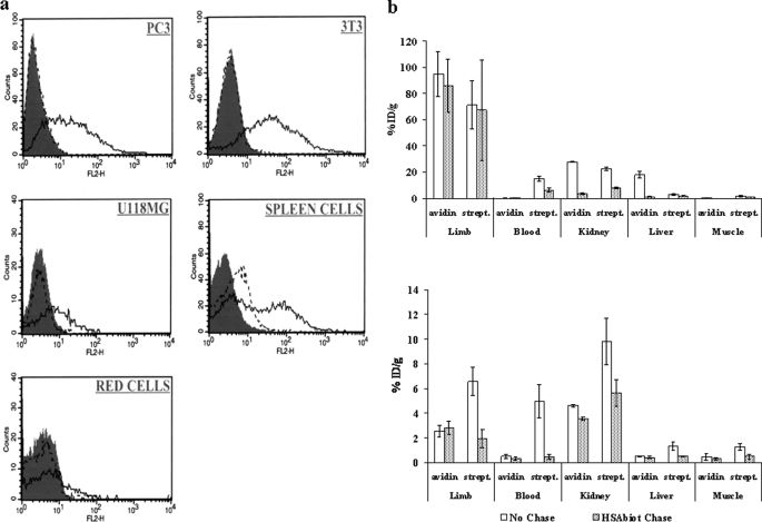
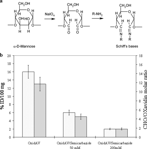
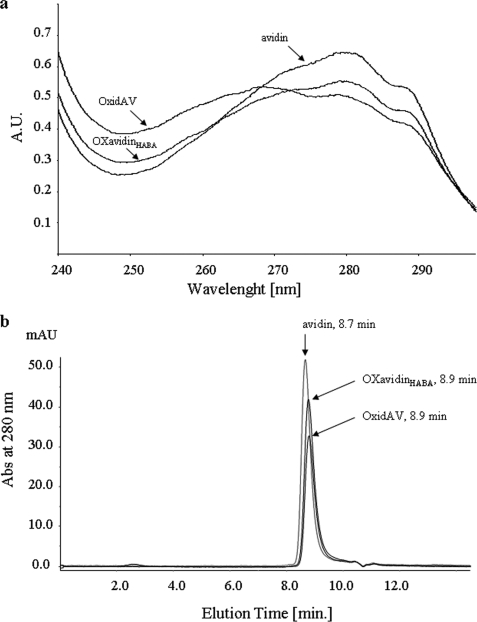
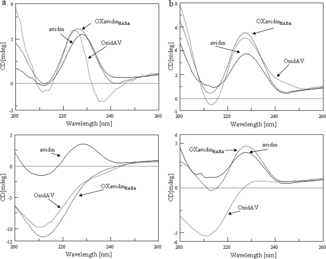
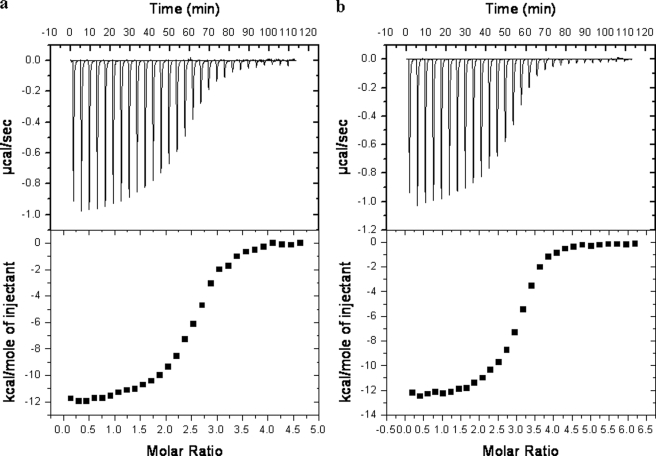
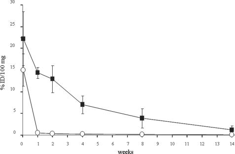
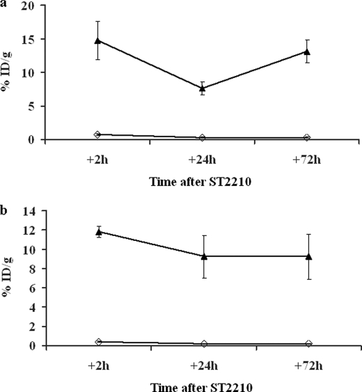
Chicken avidin and bacterial streptavidin are widely employed in vitro for their capacity to bind biotin, but their pharmacokinetics and immunological properties are not always optimal, thereby limiting their use in medical treatments. Here we investigate the biochemical and biological properties of a new modified avidin, obtained by ligand-assisted sodium periodate oxidation of avidin. This method allows protection of biotin-binding sites of avidin from inactivation caused by the oxidation step and delay of avidin clearance from injected tissue by generation of aldehyde groups from avidin carbohydrate moieties. Oxidized avidin shows spectroscopic properties similar to that of native avidin, indicating that tryptophan residues are spared from oxidation damage. In strict agreement with these results, circular dichroism and isothermal titration calorimetry analyses confirm that the ligand-assisted oxidation preserves the avidin protein structure and its biotin binding capacity. In vitro cell binding and in vivo tissue residence experiments demonstrate that aldehyde groups provide oxidized avidin the property to bind cellular and interstitial protein amino groups through Schiff's base formation, resulting in a tissue half-life of 2 weeks, compared with 2 h of native avidin. In addition, the efficient uptake of the intravenously injected (111)In-BiotinDOTA (ST2210) in the site previously treated with modified avidin underlines that tissue-bound oxidized avidin retains its biotin binding capacity in vivo. The results presented here indicate that oxidized avidin could be employed to create a stable artificial receptor in diseased tissues for the targeting of biotinylated therapeutics.


Figures

FIGURE 1.
FIGURE 1.
In vitro cell binding and in vivo uptake of 111In-ST2210 by avidin and streptavidin. a, flow cytometry of human prostate cancer PC3, mouse fibroblast NIH3T3, human glioma U118MG, mouse spleen, and red blood cells. The cells were incubated at 37 °C for 2 h in the presence of avidin (solid line) or streptavidin (dotted line) and the binding detected by biotin-B-PE. The gray peaks represent the background. b, uptake of intravenously administered 111In-ST2210 by avidin or streptavidin (strept.) 1 (top panel) or 24 (bottom panel) h after their intramuscular injection in a limb with or without HSAbiot chase. The mice were sacrificed 1 h after 111In-ST2210 injection. The highest uptake of radiolabeled biotin is evident in the treated limb at 1 h but not at 24 h. HSAbiot chase reduces background in nontarget organs. Uptake in controlateral untreated limbs is also indicated (muscle). The data are the averages of five mice, and the bars represent the standard deviations.
FIGURE 2.
FIGURE 2.
a, chemical strategy for oligosaccharide oxidation by sodium periodate and Schiff's base formation. b, dependence of OxidAV tissue residence on reactive aldehyde groups generated by oxidation with sodium periodate. Tissue residence of oxidized avidin (white) is reduced by reduction of its number of CHO groups (gray) with 50 or 200 mm semicarbazide. Evaluation was performed 24 h after injection.
FIGURE 3.
FIGURE 3.
a, absorption spectra of OxidAV and OXavidinHABA, compared with that of the unmodified avidin, in the UV range 240–300 nm. The UV spectrum of OXavidinHABA is similar to that of avidin, whereas that of OxidAV exhibits increased absorbance in the 250–260 nm region, caused by the formation on tryptophan of a substituted oxindole. b, size exclusion chromatography analysis of avidin, OxidAV and OXavidinHABA on a BiosepSEC S3000 column. Peaks of oxidized avidins appear slightly delayed compared with the one of avidin, most likely because of modification of hydrodynamic behavior consequent to oligosaccharides oxidation.
FIGURE 4.
FIGURE 4.
Circular dichroism spectra of native and oxidized avidins. a, CD spectra, recorded at 25 °C (top panel), show that OxidAV, and OXavidinHABA lack their secondary structure when heated from 25 to 95 °C (bottom panel), if compared with avidin. b, the addition of d-biotin stabilizes OXavidinHABA and avidin upon thermal denaturation (bottom panel) unlike OxidAV.
FIGURE 5.
FIGURE 5.
Calorimetric titration of OXavidinHABA (a, top panel) and avidin (b, top panel) with ST2210 in sodium acetate 100 mm, pH 5.5. The heat of dilution of ST2210 into buffer was subtracted. Bottom panels, enthalpy/mole of ST2210 injected versus ST2210/OXavidinHABA and avidin molar ratio, respectively.
FIGURE 6.
FIGURE 6.
Long term kinetics of 125I-labeled avidin (○) and OXavidinHABA (■) after intramuscular injection in the limb of Balb/c mice. At the indicated time points the mice were sacrificed, and samples of treated limb were weighed and counted in a γ counter. The data are expressed as the percentages of injected dose/100 mg (% ID/100 mg) of tissue. Each point corresponds to the average of five mice. The bars represent standard deviations.
FIGURE 7.
FIGURE 7.
Uptake of 111In-ST2210 in the muscular limb pretreated 16 (a) or 48 (b) h earlier with avidin (◇) or OXavidinHABA (▴). The mice were sacrificed 2, 24, or 72 h after intravenous injection of 111In-ST2210, and samples of treated limb were weighed and counted in a γ counter. The data are expressed as the percentages of injected dose/g (% ID/g) of tissue. Each point is the average of five mice. The bars represent standard deviations. This result confirms OXavidinHABA tissue stability and shows its capacity to uptake and keep bound the radioactive biotin.

Similar articles

Cited by

References

    1. Green N. M. (1990) Methods Enzymol. 184, 51–67 - PubMed
    1. Wilchek M., Bayer E. A. (1990) Methods Enzymol. 184, 5–13 - PubMed
    1. Hnatowich D. J., Virzi F., Rusckowski M. (1987) J. Nucl. Med. 28, 1294–1302 - PubMed
    1. Sakahara H., Saga T. (1999) Adv. Drug Deliv. Rev. 37, 89–101 - PubMed
    1. Oehr P., Westermann J., Biersack H. J. (1988) J. Nucl. Med. 29, 728–729 - PubMed

LinkOut - more resources