. 2010 Apr;30(7):1582-92.
doi: 10.1128/MCB.01605-09. Epub 2010 Jan 25.

The SAM domains of Anks family proteins are critically involved in modulating the degradation of EphA receptors

Affiliations

The SAM domains of Anks family proteins are critically involved in modulating the degradation of EphA receptors

Jieun Kim et al. Mol Cell Biol. 2010 Apr.

Abstract

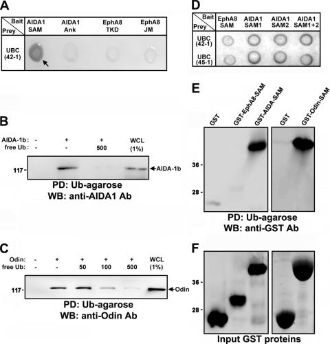
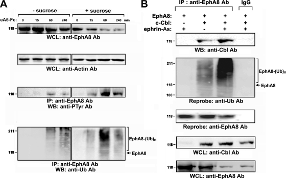
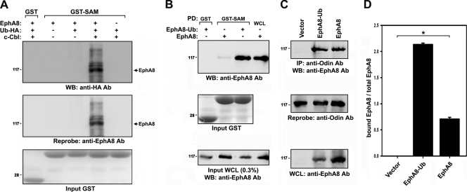
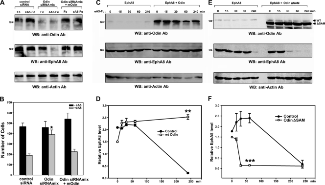
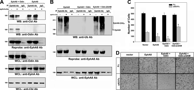
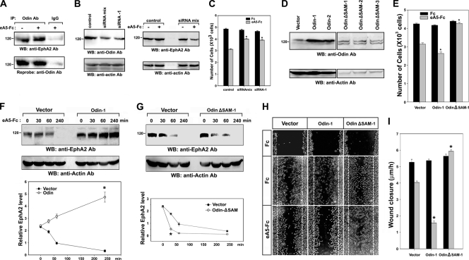
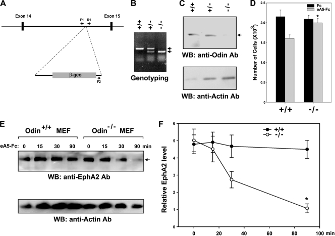
We recently reported that the phosphotyrosine-binding (PTB) domain of Anks family proteins binds to EphA8, thereby positively regulating EphA8-mediated signaling pathways. In the current study, we identified a potential role for the SAM domains of Anks family proteins in EphA signaling. We found that SAM domains of Anks family proteins directly bind to ubiquitin, suggesting that Anks proteins regulate the degradation of ubiquitinated EphA receptors. Consistent with the role of Cbl ubiquitin ligases in the degradation of Eph receptors, our results revealed that the ubiquitin ligase c-Cbl induced the ubiquitination and degradation of EphA8 upon ligand binding. Ubiquitinated EphA8 also bound to the SAM domains of Odin, a member of the Anks family proteins. More importantly, the overexpression of wild-type Odin protected EphA8 and EphA2 from undergoing degradation following ligand stimulation and promoted EphA-mediated inhibition of cell migration. In contrast, a SAM domain deletion mutant of Odin strongly impaired the function of endogenous Odin, suggesting that the mutant functions in a dominant-negative manner. An analysis of Odin-deficient primary embryonic fibroblasts indicated that Odin levels play a critical role in regulating the stability of EphA2 in response to ligand stimulation. Taken together, our studies suggest that the SAM domains of Anks family proteins play a pivotal role in enhancing the stability of EphA receptors by modulating the ubiquitination process.


Figures

FIG. 1.
FIG. 1.
Yeast two-hybrid screening showing that ubiquitin interacts with AIDA1-SAM domains. (A) Analysis of the binding of AIDA1-SAM domains to ubiquitin by X-Gal staining. As negative controls, the ankyrin (Ank) repeats of AIDA-1b, the tyrosine kinase domain (TKD) of EphA8, and the JM domain of EphA8 served as bait. UBC42-1 is a clone identified through library screening that contained part of human ubiquitin chain gene (UBC). (B) Pulldown (PD) assay using Ub-agarose beads to probe lysates of HEK293 cells expressing full-length AIDA-1b. Free ubiquitin (500 nM) was used as a competitor for binding to Ub-agarose. WB analysis was carried out to determine the level of AIDA1 bound to Ub-agarose. Ab, antibody. (C) Experiments were performed as described for panel B, except that cell lysates from Odin-expressing cells were used. WCL, whole-cell lysate. (D) A yeast two-hybrid assay was performed as described for panel A. SAM1 and SAM2 indicate the isolated SAM domains of AIDA-1. UBC45-1 represents a second UBC clone identified through library screening. (E) Ub-agarose pulldown assay of SAM domains fused to GST. (F) The samples from panel E were examined by Coomassie blue staining to determine the levels of GST fusion protein.
FIG. 2.
FIG. 2.
Cbl E3 ligase mediates ubiquitination and degradation of EphA8. (A) HEK293T cells were transfected with an expression vector for EphA8 for 24 h and then treated with sucrose for 30 min. Cells were stimulated with preclustered ephrin-A5-Fc (2 μg/ml) for the indicated times. Cell extracts were subjected to immunoprecipitation using an anti-EphA8 antibody, and immune complexes were analyzed by WB using antiphosphotyrosine (PTyr) or antiubiquitin (Ub) antibodies, as indicated. Whole-cell lysates (WCL) were directly examined by WB to determine the levels of the indicated antigens. (B) HEK293T cells were cotransfected with an expression vector for EphA8 (1 μg) and an expression vector for c-Cbl (2 μg) and then treated with NIH 3T3 cells (express endogenous ephrin-A ligands) for 30 min.
FIG. 3.
FIG. 3.
The SAM domains of Anks family proteins associate with the ubiquitinated EphA8 receptor. (A) GST pulldown assay of ubiquitinated EphA8 using AIDA-1 SAM domains. Transfections were performed as described for Fig. 2B, except that an expression vector for HA-tagged ubiquitin (1 μg) was used. The arrow indicates the position of nonubiquitinated EphA8. (B) GST pulldown assay of EphA8-Ub using AIDA-1 SAM domains. Note that expression of EphA8-Ub was lower than that of wild-type EphA8 (third panel). (C) HEK293T cells were transfected with the indicated expression constructs. Cell lysates were subjected to immunoprecipitation using an anti-Odin antibody, and immune complexes were analyzed by WB using an anti-EphA8 antibody (top panel). The blot was reprobed to determine the levels of endogenous Odin (second panel). WCL were directly probed by WB to determine the levels of EphA8 and EphA8-Ub (third panel). (D) The data in panel C were quantitated, and the amount of EphA8 in association with Odin (panel C, top panel) was normalized to total EphA8 (panel C, third panel). Data represents the means ± standard errors (SE) from three independent experiments. EphA8-Ub associated with Odin more strongly than wild-type EphA8 (*, P < 0.001 by analysis of variance [ANOVA]).
FIG. 4.
FIG. 4.
The SAM domains of Odin are required to protect EphA8 from degradation. (A) HEK293T cells were transfected with an expression vector for EphA8 and then retransfected with control siRNA or human Odin siRNA (SMART pool). Mouse Odin cDNA expression vector was also cotransfected with human Odin siRNA for the rescue experiment. At 48 hours after transfection, cells were treated with cycloheximide for 30 min and then incubated with preclustered ephrin-A5-Fc for 1 h. WCL were analyzed by WB using antibodies specific for the indicated antigens. (B) Inhibition of cell migration by ephrin-A5-stimulated EphA8 requires Odin. HEK293T cells were transfected as described for panel A. Cells were then allowed to migrate toward the lower compartment of a Boyden chamber for 4 h. Data represent the mean ± SE from three independent experiments. Cell migration under different conditions was compared with that under the control siRNA-transfected and ephrin-A5-stimulated conditions (*, P < 0.001 by ANOVA). (C) HEK293T cells were cotransfected with an expression vector for EphA8 and a control or Odin expression vector for 48 h. Cells were treated with cycloheximide for 30 min and then stimulated with preclustered ephrin-A5-Fc for the indicated times. (D) The data in panel C were quantitated, and the levels of EphA8 were normalized to the actin content. Data represent the mean ± SE from three independent experiments. The x axis represents the stimulation time. **, P < 0.01 compared to cells expressing EphA8 alone at the 4-h time point (Student's t test). (E and F) Experiments were carried out as described for panels C and D, except that an expression vector for a SAM deletion mutant of Odin, not wild-type Odin, was used. ***, P < 0.01 compared to cells expressing EphA8 alone at the 1-h time point (Student's t test).
FIG. 5.
FIG. 5.
The SAM domain of Odin interferes with the ubiquitination of EphA8. (A) HEK293T cells were transfected with an expression vector for EphA8 together with a control or Odin expression vector for 48 h and then treated with NIH 3T3 cells for 4 h. Cell lysates were subjected to immunoprecipitation using an anti-EphA8 antibody, and immune complexes were analyzed by WB using antibodies specific for Cbl (top panel) or Odin (second panel). Increasing the amount of cell lysate used for immunoprecipitation (lanes 4 and 5) resulted in detectable EphA8 in complex with endogenous Odin (data not shown). WCL were also analyzed by WB using antibodies for the indicated antigens. (B) Experiments were performed as described for Fig. 5A, except that immune complexes were analyzed by WB using an anti-Ub antibody. (C and D) Inhibition of cell migration by ephrin-A5-stimulated EphA8 requires the SAM domains of Odin. HEK293T cells were transfected as described for panel A. Cells were then allowed to migrate toward the lower compartment of a Boyden chamber for 4 h. Data represents the means ± SE from three independent experiments. The inhibitory effect of ephrin-A5 on cell migration under different conditions was compared with that in the control vector-transfected condition (*, P < 0.01 by ANOVA).
FIG. 6.
FIG. 6.
The SAM domains of Odin are required to protect EphA2 from degradation in MDA-MB-231 cells. (A) MDA-MB-231 cell lysates were subjected to immunoprecipitation using an anti-Odin antibody, and immune complexes were analyzed by WB using an anti-EphA2 antibody (top panel). The blot was reprobed to determine the levels of endogenous Odin (second panel). (B) MDA-MB-231 cells were transfected with a control siRNA, SMART pool siRNAs (siRNA mix), or a specific siRNA. At 48 hours after transfection, 20% of cells were directly analyzed by WB using antibodies specific for the indicated antigens (left panels), whereas the rest of cells were treated with cycloheximide for 30 min and then incubated with preclustered ephrin-A5-Fc for 1 h (right panels). (C) MDA-MB-231 cells were transfected as described for panel B. Cells were then allowed to migrate toward the lower compartment of a Boyden chamber for 4 h. Data represents the means ± SE from three independent experiments. Cell migration under different conditions was compared with that under the control siRNA-transfected and ephrin-A5-stimulated condition (*, P < 0.001 by ANOVA). (D) MDA-MB-231 cells were stably transfected with an expression vector for wild-type Odin or a SAM deletion mutant of Odin. Cell lysates were directly analyzed by WB using antibodies specific for Odin (top panels) or actin (bottom panels). (E) Cell migration experiments were performed as described for panel C. Cell migration under different conditions was compared with that in the vector-transfected and ephrin-A5-stimulated condition (*, P < 0.01 by ANOVA). (F) Experiments were performed as described for Fig. 4C, except MDA-MB-231 cell lysates were analyzed by WB using an anti-EphA2 antibody. *, P < 0.01 compared to cells expressing vector alone at the 4-h time point (Student's t test). (G) Experiments were performed as described for Fig. 4E, except MDA-MB-231 cell lysates were analyzed by WB using an anti-EphA2 antibody. *, P < 0.05 compared to cells expressing vector alone at the 30-min time point (Student's t test). (H and I) Wound-healing assays were carried out using cells that were prepared as described for panels F and G. The wound closure rate (y axis) was calculated from the average distance that cells at the wound edge migrated from their starting point over a period of 6 h. Data represent the means ± SE from three independent experiments. Wound closure rates under different conditions were compared with those in the vector-transfected and ephrin-A5-stimulated conditions (*, P < 0.01 by ANOVA).
FIG. 7.
FIG. 7.
Odin is required to protect EphA2 from degradation in MEFs. (A) Schematic representation of the vector integration site within the 14th intron of the mouse Odin gene in the ES clone CF0537. Exons are shown as black boxes, and the locations of the PCR primers (F1, F2, and R1) used in genotyping are indicated. (B) A typical genotyping analysis showing the 479-bp PCR fragment for the wild-type allele and the 310-bp product for the mutant allele. (C) Fibroblasts were derived from 13.5-day embryos, and the expression of Odin was analyzed by WB. (D) MEFs derived from Odin−/− knockout and Odin+/+ wild-type embryos were allowed to migrate toward the lower compartment of a Boyden chamber for 2 h. Data represents the means ± SE from three independent experiments. *, P < 0.01 compared to Odin+/+ cells (Student's t test). (E and F) Experiments were performed as described for Fig. 4C and E, except MEF lysates were analyzed by WB using an anti-EphA2 antibody. *, P < 0.01 compared to Odin+/+ cells at the 90-min time point (Student's t test).

References

    1. Arvanitis, D., and A. Davy. 2008. Eph/ephrin signaling: networks. Genes Dev. 22:416-429. - PMC - PubMed
    1. Batlle, E., J. Bacani, H. Begthel, S. Jonkheer, A. Gregorieff, M. van de Born, N. Malats, E. Sancho, E. Boon, T. Pawson, S. Gallinger, S. Pals, and H. Clevers. 2005. EphB receptor activity suppresses colorectal cancer progression. Nature 435:1126-1130. - PubMed
    1. Brantley-Sieders, D. M., G. Zhuang, D. Hicks, W. B. Fang, Y. Hwang, J. M. Cates, K. Coffman, D. Jackson, E. Bruckheimer, R. S. Muraoka-Cook, and J. Chen. 2008. The receptor tyrosine kinase EphA2 promotes mammary adenocarcinoma tumorigenesis and metastatic progression in mice by amplifying ErbB2 signaling. J. Clin. Invest. 118:64-78. - PMC - PubMed
    1. Chen, J., G. Zhuang, L. Frieden, and W. Debinski. 2008. Eph receptors and Ephrins in cancer: common themes and controversies. Cancer Res. 68:10031-10033. - PMC - PubMed
    1. Cheng, N., D. M. Brantley, H. Liu, Q. Lin, M. Enriquez, N. Gale, G. Yancopoulos, D. P. Cerretti, T. O. Daniel, and J. Chen. 2002. Blockade of EphA receptor tyrosine kinase activation inhibits vascular endothelial cell growth factor-induced angiogenesis. Mol. Cancer Res. 1:2-11. - PubMed

Publication types

MeSH terms