. 2010 Apr;30(7):1634-49.
doi: 10.1128/MCB.01164-09. Epub 2010 Jan 25.

A novel mechanism of sequestering fibroblast growth factor 2 by glypican in lipid rafts, allowing skeletal muscle differentiation

Affiliations

A novel mechanism of sequestering fibroblast growth factor 2 by glypican in lipid rafts, allowing skeletal muscle differentiation

Jaime Gutiérrez et al. Mol Cell Biol. 2010 Apr.

Abstract

Heparan sulfate proteoglycans (HSPGs) are critical modulators of growth factor activities. Skeletal muscle differentiation is strongly inhibited by fibroblast growth factor 2 (FGF-2). We have shown that HSPGs present at the plasma membrane are expressed in myoblasts and are downregulated during muscle differentiation. An exception is glypican-1, which is present throughout the myogenic process. Myoblasts that do not express glypican-1 exhibit defective differentiation, with an increase in the receptor binding of FGF-2, concomitant with increased signaling. Glypican-1-deficient myoblasts show decreased expression of myogenin, the master gene that controls myogenesis, myosin, and the myoblast fusion index. Reversion of these defects was induced by expression of rat glypican-1. Glypican-1 is the only HSPG localized in lipid raft domains in myoblasts, resulting in the sequestration of FGF-2 away from FGF-2 receptors (FGFRs) located in nonraft domains. A chimeric glypican-1, containing syndecan-1 transmembrane and cytoplasmic domains, is located in nonraft domains interacting with FGFR-IV- and enhanced FGF-2-dependent signaling. Thus, glypican-1 acts as a positive regulator of muscle differentiation by sequestering FGF-2 in lipid rafts and preventing its binding and dependent signaling.


Figures

FIG. 1.
FIG. 1.
Glypican-1 is required for a successful muscular differentiation process. (A) C2C12 myoblasts (WT) were infected with a lentiviral vector to generate a stable clone that expresses an shRNA control (shCtrl) or an shRNA specific for mouse glypican-1 (C6). Glypican-1 levels were determined by Western blot analysis using anti-stub antibodies that recognize a neoepitope generated in the heparan sulfate chains after digestion with Hase, enabling the core proteins of any HSPG to be visualized. syn-3, syn-1, syn-2, and syn-4 represent syndecan-3, -1, -2, and -4, respectively. (B) WT, shCtrl, and C6 myoblasts were induced to differentiate for 0, 2, 4, and 6 days in the differentiation medium (Days DM). Cell extracts were analyzed by Western blotting for myogenin, myosin. and caveolin-3. Tubulin levels are indicated as a loading control. In A and B, the molecular weights are indicated in thousands. (C) In a parallel experiment, WT and C6 myoblasts were fixed and analyzed by phase contrast microscopy or indirect immunofluorescence for glypican-1 (red) and myosin (green) after 5 days of differentiation (Myotubes D5). Nuclei were subjected to Hoechst staining (blue).
FIG. 2.
FIG. 2.
Reexpression of glypican-1 restores the impaired muscular differentiation observed in glypican-1-deficient myoblasts. (A) WT and C6 myoblasts were transiently transfected with shGly and with rat glypican-1 (Gly), respectively. At 48 h after transfection, the myoblasts were induced to differentiate into myotubes for 4 days (Myotubes D4). The extracts were analyzed by immunoblotting for the late muscle differentiation markers, myosin, and Cav-3. GAPDH levels were used as a loading control. (B) Phase contrast images of each experimental condition of A, at day 4 of differentiation. (C) The glypican-1 protein levels of the myoblast transfected as described in the Fig. 1A legend were determined after 48 h by immunoblot analysis using the anti-stub, as described in the same legend. (D) The glypican-1 protein levels of the myoblast transfected as described for panel A were determined after 48 h by immunoblot analysis with a glypican-1-specific antibody.
FIG. 3.
FIG. 3.
The binding of FGF-2 to its receptors is augmented in glypican-1-deficient myoblasts. (A) FGF-2 cell surface receptors of WT myoblasts transiently transfected with or without shCtrl and shGly, and of C6 myoblasts transiently transfected with or without rat glypican-1 (C6-Gly), were affinity cross-linked to 125I-FGF-2 at 4°C. Cell extracts were separated on SDS-PAGE and then exposed to a phosphorimager (left). As shown on the right, the gel was stained with Coomassie blue as a loading control. (B) The same extracts described for panel A (left) were analyzed by Western blotting to determine the total protein levels of FGFR-I and FGFR-IV. GAPDH levels were used as a loading control. (C) Myoblasts were treated with or without Hase, and then FGFRs were affinity cross-linked to 125I-FGF-2 at 4°C in the presence or absence of an excess of cold FGF-2. As shown on the right, the gel was stained with Coomassie blue as a loading control.
FIG. 4.
FIG. 4.
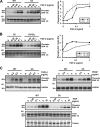
Myoblasts deficient in glypican-1 are more sensitive to FGF-2 but not to other heparin binding growth factors. (A) WT and C6 myoblasts were treated with the indicated concentrations of FGF-2 for 5 min. Cell extracts were analyzed for phospho-ERK1/2 by immunoblotting. The levels of total ERK1/2 were used as a loading control. On the right, a quantification of two independent experiments is shown. (B) C6 myoblasts were transiently transfected with or without rat glypican-1 (C6-Gly), and at 48 h after transfection, the cells were treated as described for panel A. A quantification of this experiment is shown on the right. (C) WT and C6 myoblasts were treated with the indicated concentrations of TGF-β-1 and PDGF for 15 min or HGF for 5 min. The levels of phospho-Smad 2 or phospho-ERK1/2 were determined by immunoblot analysis. GAPDH or total ERK1/2 levels were used as a loading control.
FIG. 5.
FIG. 5.
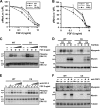
The absence of glypican-1 induces an increase in the FGF-2-dependent inhibition of the muscular differentiation process. (A) WT and C6 myoblasts were transiently cotransfected with pMyo-luc, the transfection control plasmid (pRL-SV40), an empty pcDNA3.1 plasmid as a control, or rat glypican-1 to reexpress glypican-1 in the C6 myoblasts (C6-Gly). At 48 h after transfection, the cells were induced to differentiate for 30 h in the presence of FGF-2 at the indicated concentrations. The values (pMyo-Luc/pRL-SV40 activity) are expressed as percentages of reporter activity in the absence of FGF-2 for each set of experimental conditions. (B) WT myoblasts were cotransfected with the reporter system as described for panel A and with shCtrl or shGly. After 30 h in the differentiation medium, pMyo-Luc and pRL-SV40 activities were determined and are expressed as described for panel A. The values shown in panels A and B are the results obtained from three independent experiments performed in triplicate and correspond to the mean and standard deviations. (C and E) WT or C6 myoblasts were treated with FGF-2 preincubated with or without increasing concentrations of a soluble form of the FGFR-I [FGFR(S)] or a neutralizing antibody against FGF-2 (anti-FGF-2), respectively. The phospho- and total ERK1/2 levels were determined by immunoblot analysis. (D and F) WT and C6 myoblasts were induced to differentiate in the presence or absence of the FGFR(S) and the anti-FGF-2, respectively. The anti-FGF-2 was replaced daily. Myosin and myogenin levels were analyzed by immunoblotting after 2 or 4 days. Tubulin levels were used as a loading control in both cases.
FIG. 6.
FIG. 6.
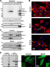
Glypican-1 is the only HSPG associated with lipid rafts. (A) C2C12 myoblasts were lysed and then fractionated in sucrose density gradients (5 to 45%). The 12 fractions collected were analyzed by immunoblotting for HSPGs, as explained in the legend of Fig. 1A, as well as the lipid raft membrane protein markers, GM-1 (ganglioside GM-1),. and Cav-1. Na+/K+ATPase (ATPase), were used as a nonlipid raft domain marker. On the left, the molecular weight standards are indicated in thousands. (B) Indirect immunocytolocalization analysis for glypican-1 (red) in C2C12 myoblasts treated with or without MβCD or PI-PLC. The nuclei were subjected to Hoechst staining (blue). The arrows indicate the punctuated pattern of glypican-1 on the cell surface, and the arrowheads point at the ECM-associated glypican-1. (C) C2C12 myoblasts were treated with MβCD or left untreated and then fractionated as described for panel A. The fractions were analyzed for glypican-1 and Cav-1 distribution by Western blotting. (D) As shown on the left, C6 clone myoblasts were transiently transfected with rat glypican-1 containing a FLAG epitope in its amino terminal (F-Gly) or the empty vector. After 48 h, the cells were fractionated as described for panel A. The 12 fractions were pooled into three groups: group I (fractions 1 to 4), group II (fractions 5 to 8), and group III (fractions 9 to 12). The fractions in each group were analyzed for the distribution of rat glypican-1 by using an anti-FLAG antibody. As shown on the right, in a parallel experiment, C6 myoblasts transfected with F-Gly were treated with MβCD or left untreated and then fixed and analyzed by immunofluorescence for the presence of the FLAG epitope. syn-3, syn-1, syn-2, and syn-4 represent syndecan-3, -1, -2, and -4, respectively.
FIG. 7.
FIG. 7.
Glypican-1 concentrates FGF-2 in lipid raft microdomains that exclude FGF-2 signaling receptors. (A) WT and C6 myoblasts were incubated with [125I]-FGF-2 for 3 h at 4°C and then fractionated as described for Fig. 1. The fractions were analyzed for HSPGs by using anti-stub, as described for Fig. 1A, or exposed to a phosphorimager to detect the distribution of 125I-FGF-2. (B) C2C12 myoblasts were fractionated as explained for Fig. 7A. The fractions were analyzed for the distribution of FGFR-I and FGFR-IV as well as membrane distribution markers Cav-1, GM-1, and Na+/K+ATPase (ATPase). (C) FGF-2 cell surface receptors in C2C12 myoblasts were affinity cross-linked to 125I-FGF-2 at 4°C. The cells were lysed and fractionated as explained for Fig. 7A. Aliquots of each fraction were separated on SDS-PAGE (4 to 10%). The gel was dried and exposed to a phosphorimager (upper panel) or analyzed by Western immunoblotting for Cav-1, GM-1, and ATPase. 125I-FGF-2-FGFR-I and 125I-FGF-2-FGFR-IV correspond to 125I-FGF-2 cross-linked to FGFR-I and FGFR-IV, respectively.
FIG. 8.
FIG. 8.
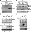
Expression of glypican-1 outside the lipid raft acts like that of a FGF-2 coreceptor. (A) C6 myoblasts were transiently transfected with a chimeric HSPG (F-GlySyn) composed of the extracellular domain of rat glypican-1 and the transmembrane and cytosolic domain of mouse syndecan-1, containing a FLAG epitope. After 48 h, the cells were lysed and fractionated as described for Fig. 6A. The distribution of F-GlySyn was evaluated by anti-FLAG analysis. (B) C6 myoblasts were transfected with or without rat glypican (F-Gly) or F-GlySyn and treated with increasing concentrations of FGF-2. The phospho-ERK1/2, total ERK1/2, and tubulin levels were determined by immunoblot analysis. (C) C6 myoblasts were transfected with or without rat glypican (F-Gly) or F-GlySyn and induced to differentiate for 2 (D2) or 4 (D4) days to determine the myogenin, myosin, and tubulin levels by immunoblot analysis. (D) WT or C6 myoblasts were transiently transfected as described for panel C. After 48 h, the cells were lysed and the extracts immunoprecipitated with an anti-FLAG antibody or anti-mouse syndecan-4. The immunoprecipitates were analyzed by Western blotting for the presence of FGFR-IV, F-Gly, and F-GlySyn with an anti-anti-glypican-1 or syndecan-4.
FIG. 9.
FIG. 9.
Glypican-1 is present in the plasma membrane lipid rafts and in the extracellular matrix. (A) WT myoblasts were fractionated as shown in Fig. 7 after being washed with heparin in PBS Ca2+/Mg2+ (Heparin Wash) or PBS Ca2+/Mg2+ alone (control). The fractions were analyzed for HSPG core proteins and Cav-1 distribution. (B) Indirect immunofluorescence for glypican-1 (red) of cells treated as described for panel A. (C) Indirect immunofluorescence for glypican-1 (red) and laminin (green) in WT myoblasts.
FIG. 10.
FIG. 10.
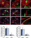
Glypican-1 is required on the plasma membrane for a proper muscular differentiation process independent from extracellular matrix-associated glypican-1. C2C12 myoblasts were transiently cotransfected with scrambled shRNA (shCtrl) and a plasmid containing the sequence for E-GFP (A, D, and I) or shGly (B, E, C, and F). At 48 h after transfection, the myoblasts were induced to differentiate for 2 days (A, B, D, and E) or 4 days (C, F, and I). The cells were fixed and analyzed by immunofluorescence for glypican-1 (red) (A, B, and C), myogenin (red) (D and E), or myosin (red) (F and I). Panels G and H present the same images as panels D and E, respectively, but without the E-GFP signal, in order to better visualize myogenin nuclear staining. The arrowheads indicate the ECM-associated glypican-1. The arrows indicate the nuclei of transfected cells. (J) Shown on the left is a quantification of the cotransfected myoblasts (shCtrl/E-GFP or shGly/E-GFP) containing myogenin-positive nuclei compared to the total cotransfected cells (E-GFP positive) after 2 days of differentiation of 10 random fields. Shown on the right is a quantification of the E-GFP-myosin-positive myotubes compared to the total number of E-GFP-expressing cells after 4 days of differentiation. The data correspond to the means ± standard errors of the results obtained with 10 random fields.

Similar articles

Cited by

References

    1. Akiyama, S., T. Katagiri, M. Namiki, N. Yamaji, N. Yamamoto, K. Miyama, H. Shibuya, N. Ueno, J. M. Wozney, and T. Suda. 1997. Constitutively active BMP type I receptors transduce BMP-2 signals without the ligand in C2C12 myoblasts. Exp. Cell Res. 235:362-369. - PubMed
    1. Anastasi, S., S. Giordano, O. Sthandier, G. Gambarotta, R. Maione, P. Comoglio, and P. Amati. 1997. A natural hepatocyte growth factor/scatter factor autocrine loop in myoblast cells and the effect of the constitutive Met kinase activation on myogenic differentiation. J. Cell Biol. 137:1057-1068. - PMC - PubMed
    1. Ashikari, S., H. Habuchi, and K. Kimata. 1995. Characterization of heparan sulfate oligosaccharides that bind to hepatocyte growth factor. J. Biol. Chem. 270:29586-29593. - PubMed
    1. Beer, C., L. Pedersen, and M. Wirth. 2005. Amphotropic murine leukaemia virus envelope protein is associated with cholesterol-rich microdomains. Virol. J. 2:36. - PMC - PubMed
    1. Bernfield, M., M. Gotte, P. W. Park, O. Reizes, M. L. Fitzgerald, J. Lincecum, and M. Zako. 1999. Functions of cell surface heparan sulfate proteoglycans. Annu. Rev. Biochem. 68:729-777. - PubMed

Publication types

MeSH terms