. 2010 Jan 25;188(2):191-8.
doi: 10.1083/jcb.200911102.

Polo-like kinase 4 kinase activity limits centrosome overduplication by autoregulating its own stability

Affiliations

Polo-like kinase 4 kinase activity limits centrosome overduplication by autoregulating its own stability

Andrew J Holland et al. J Cell Biol. .

Abstract

Accurate control of the number of centrosomes, the major microtubule-organizing centers of animal cells, is critical for the maintenance of genome integrity. Abnormalities in centrosome number can promote errors in spindle formation that lead to subsequent chromosome missegregation, and extra centrosomes are found in many cancers. Centrosomes are comprised of a pair of centrioles surrounded by amorphous pericentriolar material, and centrosome duplication is controlled by centriole replication. Polo-like kinase 4 (Plk4) plays a key role in initiating centriole duplication, and overexpression of Plk4 promotes centriole overduplication and the formation of extra centrosomes. Using chemical genetics, we show that kinase-active Plk4 is inherently unstable and targeted for degradation. Plk4 is shown to multiply self-phosphorylate within a 24-amino acid phosphodegron. Phosphorylation of multiple sites is required for Plk4 instability, indicating a requirement for a threshold level of Plk4 kinase activity to promote its own destruction. We propose that kinase-mediated, autoregulated instability of Plk4 self-limits Plk4 activity so as to prevent centrosome amplification.


Figures

Figure 1.
Figure 1.
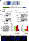
Plk4 kinase activity destabilizes the protein. (A) Schematic of mouse Plk4, showing the location of the kinase domain, the Polo-box domain (PBD), and the cryptic Polo-box domain (Swallow et al., 2005). The locations of the AS and KD mutations are also shown. (B) Immunoblot showing the expression level of WT and KD Plk4-EYFP transgenes. (C) Titration showing the relative expression level of Plk4WT and Plk4KD. (D) Immunoblot showing the expression levels of Plk4WT, Plk4AS, and Plk4KD in the presence of DMSO or 1NM-PP1. (E) Immunofluorescence images acquired 48 h after Plk4-EYFP was induced with tetracycline. Cells were grown in DMSO or 1NM-PP1. Insets depict an enlargement of centrioles. Red, SAS-6; green, Plk4-EYFP; blue, DNA. Bars, 10 µm. (F) Graph quantifying of the proportion of cells with more than four centrioles 48 h after expression of Plk4-EYFP was induced. Cells were grown in the presence of either DMSO or 1NM-PP1. Bars represent the mean of at least three independent experiments with >300 cells per condition. Error bars represent the SEM.
Figure 2.
Figure 2.
Phosphorylation of the β-TrCP–binding motif has a minor effect on the stability of mouse Plk4. (A) Cells were treated with or without the proteasome inhibitor MG132 for 8 h and immunoblotted to determine the expression level of Plk4WT and Plk4KD. (B) Expression of Plk4WT-EYFP or Plk4KD-EYFP was induced for 24 h in cells constitutively expressing Plk4KD-Myc. The expression level of the Myc and EYFP-tagged Plk4 transgenes was subsequently determined by immunoblotting. (C) The Plk4AA mutant possesses two mutations that prevent phosphorylation of the β-TrCP–binding motif (highlighted in green). (D) Immunoblot shows a modest increase in the stability of Plk4AA. (E) HEK 293 cells were cotransfected with Plk4-EYFP and Myc–β-TrCP and treated with MG132 for 4 h. Plk4-EYFP was purified using GBP-coupled beads or beads alone, and protein complexes were analyzed by immunoblotting. (F) HEK 293 cells were cotransfected with Plk4-EYFP and HA-ubiquitin and treated with MG132 for 4 h. Plk4-EYFP was purified using GBP-coupled beads or beads alone, and protein complexes were analyzed by immunoblotting.
Figure 3.
Figure 3.
Plk4 autophosphorylates a 24-aa region to promote its own destruction. (A) Schematic showing the localization of the 24-aa region that is required for Plk4 to promote its own destruction. The alignment of this region, which is rich in potential phosphorylation sites (S/T residues), is shown. This putative β-TrCP–binding motif is contained within this region and marked by a line. (B) GST-Plk4 kinase assays were performed using His-Plk4KD (aa 1–416) as a substrate. GST-Plk4WT phosphorylates itself and His-Plk4KD, but note that phosphorylation of His-Plk4KD is reduced by the Δ24 mutation. Coomassie-stained gel shows the purified protein, and the autoradiogram shows the incorporation of γ[32P]ATP. Assays were performed for 60 min, and activity was measured by scintillation counting. (C) His-Plk4 (aa 1–416) kinase assay was performed using histone H1 as a substrate. Despite the fact that WT and Δ24 His-Plk4 have similar activity toward histone H1, His-Plk4Δ24 displays reduced autophosphorylation. Coomassie-stained gel shows the purified protein, and autoradiogram shows the incorporation of γ[32P]ATP. Assays were performed for 60 min, and activity was measured by scintillation counting. (D) Immunoblot showing the relative expression levels of WT, AA, Δ24, and KD Plk4-EYFP. (E) Graph quantifying of the proportion of cells with more than four centrioles 48 h after induction of Plk4-EYFP. (F) Graph quantifying the mean number of centrioles per cell in cells with more than four centrioles. Counting was performed 48 h after induction of Plk4-EYFP. (G) Immunofluorescence images acquired 48 h after Plk4-EYFP was induced. Insets depict an enlargement of centrioles. Red, SAS-6; green, Plk4-EYFP; blue, DNA. Bars, 10 µm. C, Coomassie. Bars represent the mean of at least three independent experiments with >280 cells per condition. Error bars represent the SEM.
Figure 4.
Figure 4.
Phosphorylation of multiple sites is required for optimal Plk4 destruction. (A) Alignment showing the positions of the different phosphorylation mutations in Plk4. Immunoblot showing the relative expression levels of WT, AA, A1, A2, A3, A4, and KD Plk4-EYFP. (B) Graph quantifying of the proportion of cells with more than four centrioles 48 h after induction of Plk4-EYFP. Uninduced samples (−Tet) are shown for comparison. (C) Graph quantifying the mean number of centrioles per cell in cells with more than four centrioles. Counting was performed 48 h after induction of Plk4-EYFP. Uninduced samples are shown for comparison. (D) Immunofluorescence images acquired 48 h after Plk4-EYFP was induced. Insets depict an enlargement of centrioles. Red, SAS-6; green, Plk4-EYFP; blue, DNA. Bars, 10 µm. (E) A model for the autoregulation of Plk4 stability. Bars represent the mean of at least three independent experiments with >300 cells per condition. Error bars represent the SEM.

Similar articles

Cited by

References

    1. Basto R., Brunk K., Vinadogrova T., Peel N., Franz A., Khodjakov A., Raff J.W. 2008. Centrosome amplification can initiate tumorigenesis in flies. Cell. 133:1032–1042 10.1016/j.cell.2008.05.039 - DOI - PMC - PubMed
    1. Bettencourt-Dias M., Rodrigues-Martins A., Carpenter L., Riparbelli M., Lehmann L., Gatt M.K., Carmo N., Balloux F., Callaini G., Glover D.M. 2005. SAK/PLK4 is required for centriole duplication and flagella development. Curr. Biol. 15:2199–2207 10.1016/j.cub.2005.11.042 - DOI - PubMed
    1. Bishop A.C., Ubersax J.A., Petsch D.T., Matheos D.P., Gray N.S., Blethrow J., Shimizu E., Tsien J.Z., Schultz P.G., Rose M.D., et al. 2000. A chemical switch for inhibitor-sensitive alleles of any protein kinase. Nature. 407:395–401 10.1038/35030148 - DOI - PubMed
    1. Burkard M.E., Randall C.L., Larochelle S., Zhang C., Shokat K.M., Fisher R.P., Jallepalli P.V. 2007. Chemical genetics reveals the requirement for Polo-like kinase 1 activity in positioning RhoA and triggering cytokinesis in human cells. Proc. Natl. Acad. Sci. USA. 104:4383–4388 10.1073/pnas.0701140104 - DOI - PMC - PubMed
    1. Castellanos E., Dominguez P., Gonzalez C. 2008. Centrosome dysfunction in Drosophila neural stem cells causes tumors that are not due to genome instability. Curr. Biol. 18:1209–1214 10.1016/j.cub.2008.07.029 - DOI - PubMed

Publication types

MeSH terms

Substances