

Radiographic findings and prognosis of simple bone cysts of the jaws
- PMID: 20100916
- PMCID: PMC3520194
- DOI: 10.1259/dmfr/54872008
Radiographic findings and prognosis of simple bone cysts of the jaws
Abstract
Objective: The aim was to evaluate the possibility of radiographic prediction of the prognosis of simple bone cysts (SBCs) of the jaws.
Methods: The relationship between the radiographic findings and treatment outcome (healing or recurrence) was investigated in 31 cases treated in the authors' hospital and 108 published cases.
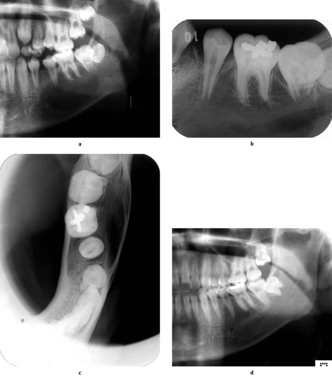
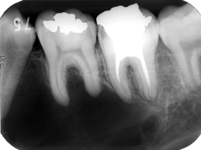
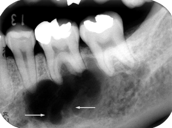
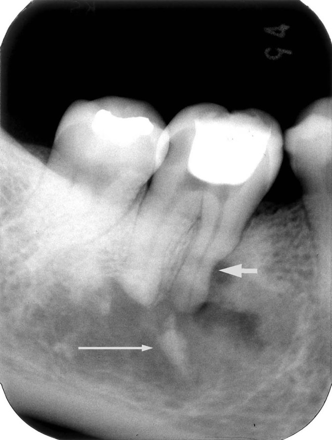
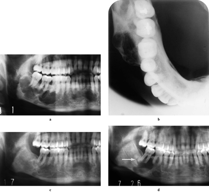
Results: In 17 of our 31 cases, the lesions had radiographic findings of a preserved lamina dura adjacent to the lesion, with a smooth margin, and no or smooth bone expansion, and all of them healed after surgery. In the other 14 cases, there was resorption of the lamina dura, a scalloped margin, nodular bone expansion, root resorption, a sclerotic mass or multiple cavities, and nine cases of recurrence. Although there was incomplete information in the published case studies, similar results were noted.
Conclusions: It was concluded that there was a relationship between the radiographic features of the lesions and prognosis. Radiographic examination should be used not only for discovering and diagnosing the lesions, but also for predicting possible prognosis.
Figures
References
-
- Weber AL, Kaneda T, Scrivani SJ, Aziz S. Jaws and temporomandibular joints. In: Som PM, Curtin HD (eds). Head and neck imaging, 4th edn. St. Louis: Mosby, 2003, pp 942–943
-
- White SC, Pharoah MJ. Oral radiology principles and interpretation, 5th edn. St. Louis: Mosby, 2004, pp 405–409.
-
- Shafer WG, Hine MK, Levy BM. A textbook of oral pathology, 4th edn. Philadelphia: WB Saunders, 1983, pp 541–544.
-
- Damante JH, Da S, Guerra EN, Ferreira Jr O. Spontaneous resolution of simple bone cysts. Dentomaxillofac Radiol 2002;31:182–186 - PubMed
-
- Sapp JP, Stark ML. Self-healing traumatic bone cysts. Oral Surg Oral Med Oral Pathol 1990;69:597–602 - PubMed
