

A comparison of two-dimensional radiography and three-dimensional computed tomography in angular cephalometric measurements
- PMID: 20100922
- PMCID: PMC3520203
- DOI: 10.1259/dmfr/82724776
A comparison of two-dimensional radiography and three-dimensional computed tomography in angular cephalometric measurements
Abstract
Objectives: The objective of this study was to assess the reliability of three-dimensional (3D) cephalometric approaches by comparing this method with authenticated traditional two-dimensional (2D) cephalometry in angular cephalometric measurements.
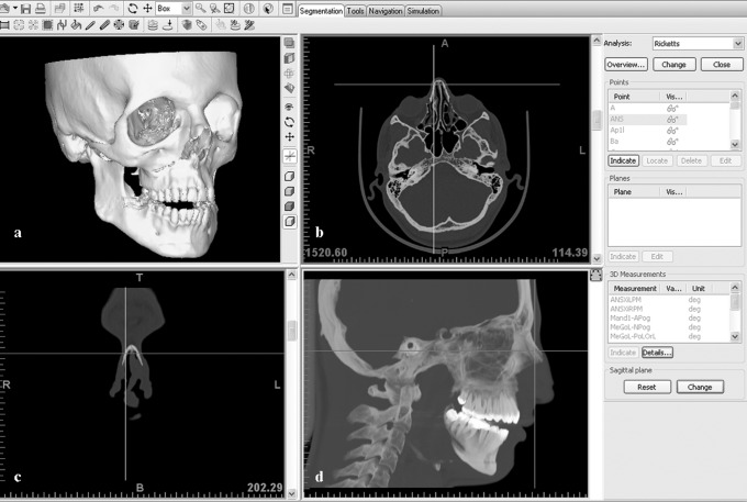
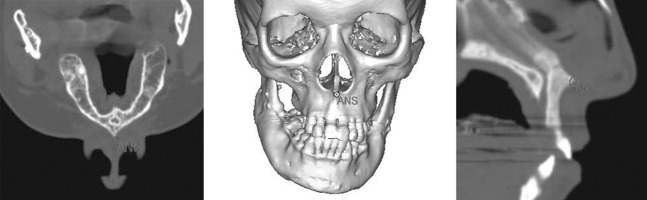
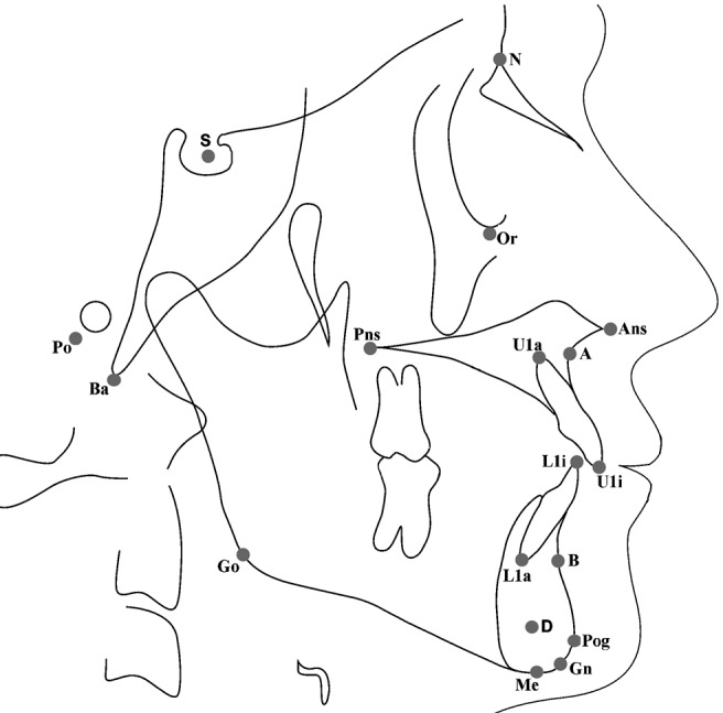
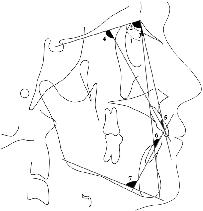
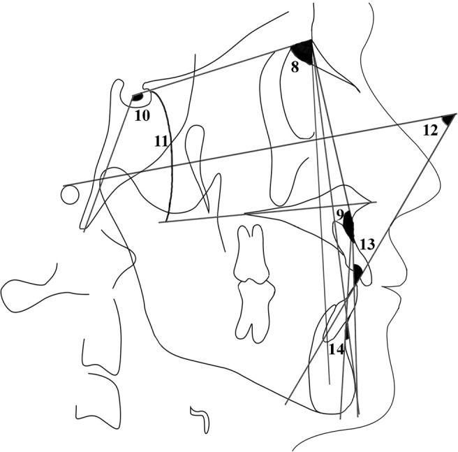
Methods: CT images and lateral cephalometric radiographs of ten patients (five women, five men) were used in this study. Raw CT data of the patients were converted to 3D images with a 3D simulation program (Mimics 9.0, Leuven, Belgium). Lateral cephalometric radiographs were used manually for 2D measurements. The comparisons of the two methods were made using 14 cephalometric angular measurements. The Wilcoxon matched-pairs signed-ranks test (alpha = 0.05) was used to determine the difference between the two methods. To assess the intra- and interobserver reproducibility, two sets of recordings made by each observer, in each modality were used. Dahlberg's formula was used to determine the intraobserver reproducibility, and the Wilcoxon matched-pairs signed-rank test (alpha = 0.05) was used to assess the interobserver reproducibility.
Results: The method errors of both observers ranged from 0.35 degrees to 0.65 degrees. In addition, there were no significant differences between the measurements of the two observers (P > 0.05). However, comparison of 2D and 3D parameters showed significant differences in U1-NA and U1-SN measurements (P < 0.05).
Conclusions: The 3D angular cephalometric analysis is a fairly reliable method, like the traditional 2D cephalometric analysis. Currently, the 3D system is likely to be more suitable for the diagnosis of cases with complex orthodontic anomalies. However, with the decrease in radiation exposure and costs in the future, 3D cephalometrics can be a suitable alternative method to 2D cephalometry.
Figures
References
-
- Broadbent BH. A new x-ray technique and its application to orthodontia. Angle Orthod 1931;51:93–114
-
- Halazonetis DJ. From 2-dimensional cephalograms to 3-dimensional computed tomography scans. Am J Orthod Dentofacial Orthop 2005;127:627–637 - PubMed
-
- Papadopoulos MA, Jannowitz C, Boettcher P, Henke J, Stolla R, Zeilhofer HF, et al. Three-dimensional fetal cephalometry: an evaluation of the reliability of cephalometric measurements based on three-dimensional CT reconstructions and on dry skulls of sheep fetuses. J Craniomaxillofac Surg 2005;33:229–237 - PubMed
-
- Moyers RE, Bookstein FL, Hunter WS. Analysis of the craniofacial skeleton: cephalometrics. In: Moyers RE (ed). Handbook of orthodontics. Chicago: Year Book Medical, 1988, pp 247–309
-
- Baumrind S, Frantz RC. The reliability of head film measurements. 1. Landmark identification. Am J Orthod 1971;60:111–127 - PubMed
