

CD72 negatively regulates KIT-mediated responses in human mast cells
- PMID: 20100931
- PMCID: PMC2824776
- DOI: 10.4049/jimmunol.0902450
CD72 negatively regulates KIT-mediated responses in human mast cells
Abstract
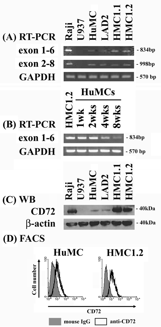
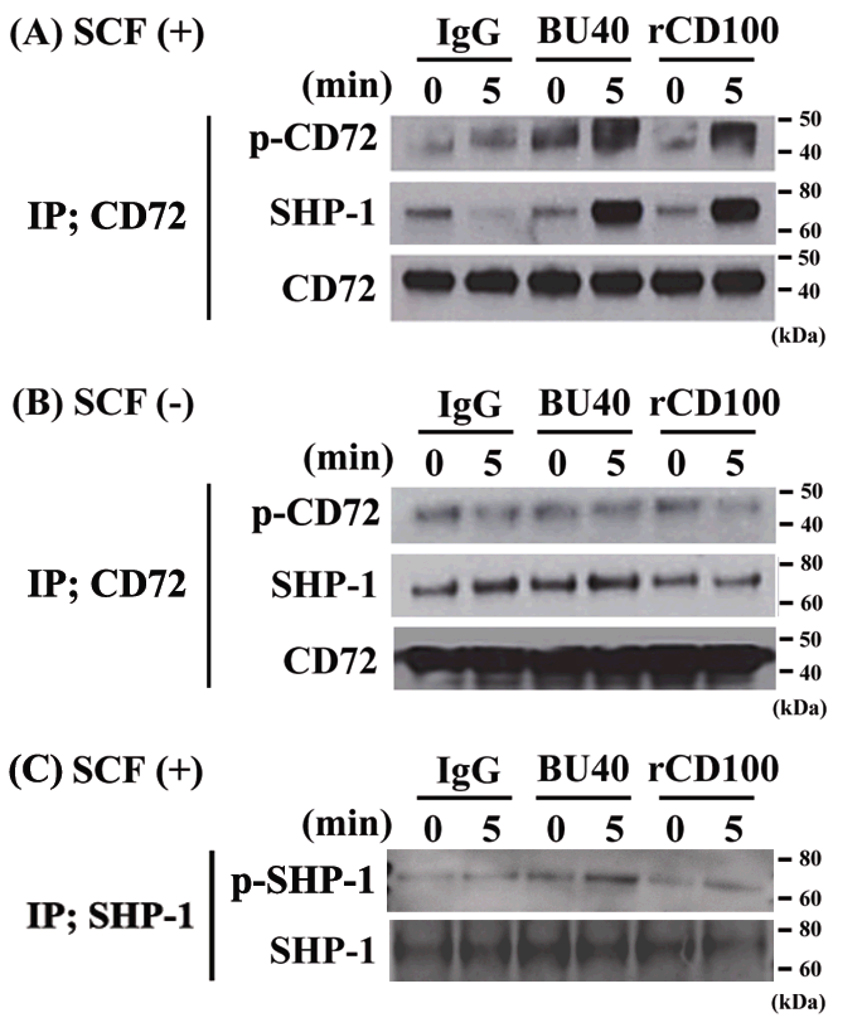
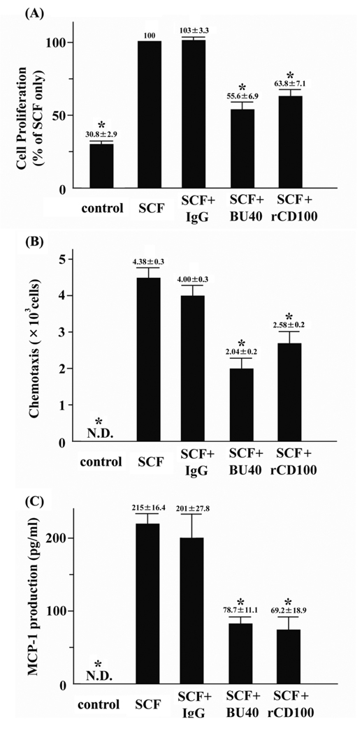
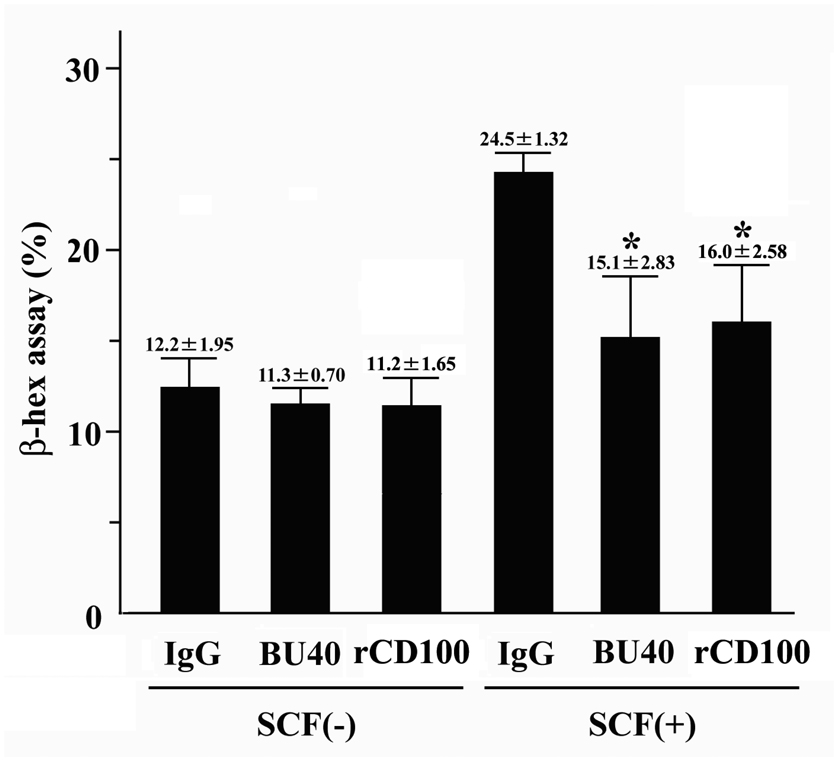
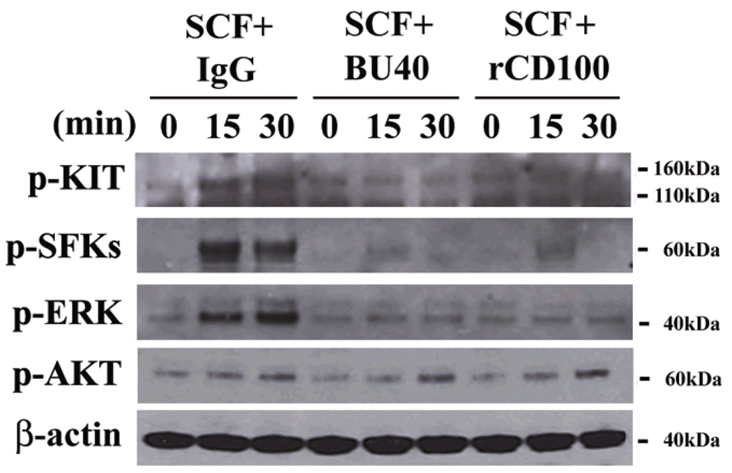
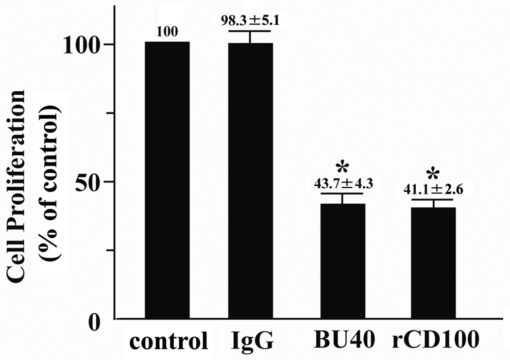
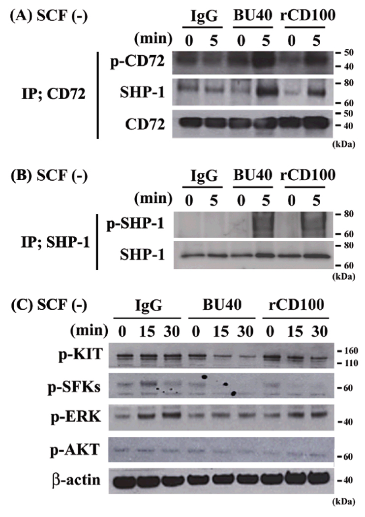
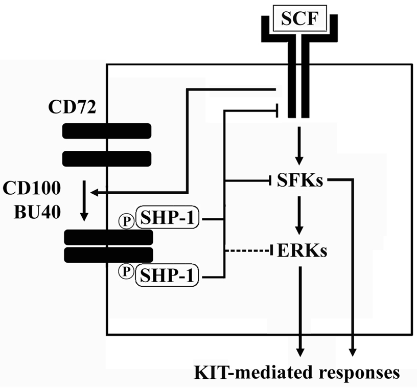
KIT activation, through binding of its ligand, stem cell factor, is crucial for normal mast cell growth, differentiation, and survival. Furthermore, KIT may also contribute to mast cell homing and cytokine generation. Activating mutations in KIT lead to the dysregulated mast cell growth associated with the myeloproliferative disorder, mastocytosis. We investigated the potential of downregulating such responses through mast cell inhibitory receptor activation. In this study, we report that the B cell-associated ITIM-containing inhibitory receptor, CD72, is expressed in human mast cells. Ligation of CD72 with the agonistic Ab, BU40, or with recombinant human CD100 (rCD100), its natural ligand, induced the phosphorylation of CD72 with a resulting increase in its association with the tyrosine phosphatase SH2 domain-containing phosphatase-1. This, in turn, resulted in an inhibition of KIT-induced phosphorylation of Src family kinases and extracellular-regulated kinases (ERK1/2). As a consequence of these effects, KIT-mediated mast cell proliferation, chemotaxis, and chemokine production were significantly reduced by BU40 and rCD100. Furthermore, BU40 and rCD100 also downregulated the growth of the HMC1.2 human mast cell line. Thus, targeting CD72 may provide a novel approach to the suppression of mast cell disease such as mastocytosis.
Figures
Similar articles
-
CD72 regulates the growth of KIT-mutated leukemia cell line Kasumi-1.Sci Rep. 2013 Oct 4;3:2861. doi: 10.1038/srep02861. Sci Rep. 2013. PMID: 24713856 Free PMC article.
-
CD72 negatively regulates mouse mast cell functions and down-regulates the expression of KIT and FcεRIα.Int Immunol. 2015 Feb;27(2):95-103. doi: 10.1093/intimm/dxu087. Epub 2014 Sep 19. Int Immunol. 2015. PMID: 25239131
-
A novel KIT-deficient mouse mast cell model for the examination of human KIT-mediated activation responses.J Immunol Methods. 2013 Apr 30;390(1-2):52-62. doi: 10.1016/j.jim.2013.01.008. Epub 2013 Jan 25. J Immunol Methods. 2013. PMID: 23357051 Free PMC article.
-
Negative regulation of mast cell proliferation by FcgammaRIIB.Mol Immunol. 2002 Sep;38(16-18):1295-9. doi: 10.1016/s0161-5890(02)00078-0. Mol Immunol. 2002. PMID: 12217398 Review.
-
Mastocytosis.Chem Immunol Allergy. 2010;95:110-124. doi: 10.1159/000315946. Epub 2010 Jun 1. Chem Immunol Allergy. 2010. PMID: 20519885 Free PMC article. Review.
Cited by
-
Regulation of mast cell responses in health and disease.Crit Rev Immunol. 2011;31(6):475-529. doi: 10.1615/critrevimmunol.v31.i6.30. Crit Rev Immunol. 2011. PMID: 22321108 Free PMC article. Review.
-
CAR-T Cells Shoot for New Targets: Novel Approaches to Boost Adoptive Cell Therapy for B Cell-Derived Malignancies.Cells. 2022 May 31;11(11):1804. doi: 10.3390/cells11111804. Cells. 2022. PMID: 35681499 Free PMC article. Review.
-
Novel identified receptors on mast cells.Front Immunol. 2012 Aug 2;3:238. doi: 10.3389/fimmu.2012.00238. eCollection 2012. Front Immunol. 2012. PMID: 22876248 Free PMC article.
-
Glycogen synthase kinase-3β is a prosurvival signal for the maintenance of human mast cell homeostasis.J Immunol. 2011 Dec 1;187(11):5587-95. doi: 10.4049/jimmunol.1101257. Epub 2011 Oct 28. J Immunol. 2011. PMID: 22039301 Free PMC article.
-
Mast cells.Curr Gastroenterol Rep. 2010 Oct;12(5):349-57. doi: 10.1007/s11894-010-0132-1. Curr Gastroenterol Rep. 2010. PMID: 20711694 Free PMC article. Review.
References
-
- Kraft S, Kinet JP. New developments in FcεRI regulation, function and inhibition. Nat. Rev. Immunol. 2007;7:365–378. - PubMed
-
- Nagata H, Worobec AS, Oh CK, Chowdhury BA, Tannenbaum S, Suzuki Y, Metcalfe DD. Identification of a point mutation in the catalytic domain of the protooncogene c-kit in peripheral blood mononuclear cells of patients who have mastocytosis with an associated hematologic disorder. Proc. Natl. Acad. Sci. USA. 1995;92:10560–10564. - PMC - PubMed
-
- Furitsu T, Tsujimura T, Tono T, Ikeda H, Kitayama H, Koshimizu U, Sugahara H, Butterfield JH, Ashman LK, Kanayama Y, Matsuzawa Y, Kitamura Y, Kanakura Y. Identification of mutations in the coding sequence of the proto-oncogene c-kit in a human mast cell leukemia cell line causing ligand-independent activation of c-kit product. J. Clin. Invest. 1993;92:1736–1744. - PMC - PubMed
Publication types
MeSH terms
Substances
Grants and funding
LinkOut - more resources
Full Text Sources
Molecular Biology Databases
Research Materials
Miscellaneous
