. 2010 Apr;184(4):1165-79.
doi: 10.1534/genetics.109.112516. Epub 2010 Jan 25.

A genomewide RNA interference screen for modifiers of aggregates formation by mutant Huntingtin in Drosophila

Affiliations

A genomewide RNA interference screen for modifiers of aggregates formation by mutant Huntingtin in Drosophila

Sheng Zhang et al. Genetics. 2010 Apr.

Abstract

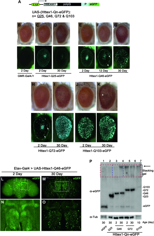
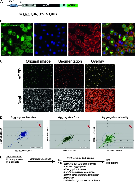
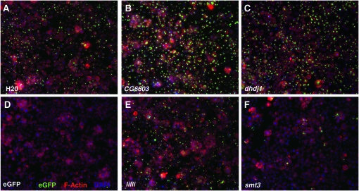
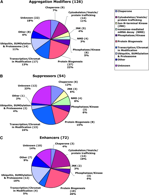
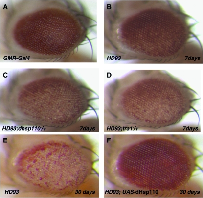
Protein aggregates are a common pathological feature of most neurodegenerative diseases (NDs). Understanding their formation and regulation will help clarify their controversial roles in disease pathogenesis. To date, there have been few systematic studies of aggregates formation in Drosophila, a model organism that has been applied extensively in modeling NDs and screening for toxicity modifiers. We generated transgenic fly lines that express enhanced-GFP-tagged mutant Huntingtin (Htt) fragments with different lengths of polyglutamine (polyQ) tract and showed that these Htt mutants develop protein aggregates in a polyQ-length- and age-dependent manner in Drosophila. To identify central regulators of protein aggregation, we further generated stable Drosophila cell lines expressing these Htt mutants and also established a cell-based quantitative assay that allows automated measurement of aggregates within cells. We then performed a genomewide RNA interference screen for regulators of mutant Htt aggregation and isolated 126 genes involved in diverse cellular processes. Interestingly, although our screen focused only on mutant Htt aggregation, several of the identified candidates were known previously as toxicity modifiers of NDs. Moreover, modulating the in vivo activity of hsp110 (CG6603) or tra1, two hits from the screen, affects neurodegeneration in a dose-dependent manner in a Drosophila model of Huntington's disease. Thus, other aggregates regulators isolated in our screen may identify additional genes involved in the protein-folding pathway and neurotoxicity.


Figures

F<sc>igure</sc> 1.—
Figure 1.—
Age- and polyQ-length-dependent formation of aggregates in Drosophila. (A) Structure of the Htt exon1 (Httex1) constructs used for the studies. eGFP-tagged Httex1 with different lengths of glutamine tract (polyQ) followed by its proline-rich region (P) are under the control of the UAS elements (Brand and Perrimon 1993). (B–P) PolyQ-length- and age-dependent formation of aggregates in Drosophila. (B–K) Httex1-Qn-eGFP were expressed in the eye using the eye-specific GMR-Gal4 driver. (Top) The general eye morphology of adults of different ages. (Bottom) Images of the same eyes under green fluorescent light, with their age and genotype indicated below. Genotypes: (B) GMR-Gal4/+ and (C–K) GMR-Gal4/+; UAS-Httex1-Qn-eGFP/+. (B) Control of GMR-Gal4 driver alone at day 2. Note that there is no visible eGFP signal in this eye (bottom) in the absence of the UAS-eGFP reporter. (C and D) Adult eyes with Httex1-Q25-eGFP expression. Q25 is found mainly in a diffuse cytoplasmic pattern at both day 2 (C) and day 30 (D). Note that the center bright spots in both eyes and in E are not aggregates but rather an optical phenomenon similar to the “deep pseudopupil,” arising from the superimposition of evenly diffuse GFP signals emitted from the underlying regularly arranged ommatidia (Franceschini 1972). (E–G) Adult eyes with Httex1-Q46-eGFP expression. At day 2 (E, left eye), Httex1-Q46 is found mainly in a diffuse cytoplasmic pattern. As the fly ages, sporadic aggregates gradually accumulate (F, white arrows) and become prominent by day 30 (G). (H and I) Adult eyes with Httex1-Q72-eGFP expression. At day 2 (H), Q72 is found mainly in a diffuse cytoplasmic pattern but sporadic aggregates are already visible (H, white arrows). Aggregates become prominent by day 30 (I). (J and K) Adult eyes with Httex1-Q103-eGFP expression. Aggregates already become prominent at day 2 (J) and persist at day 30 (K). Note also that there is a mild loss of pigment at the posterior of this eye at day 30 (K, white arrowhead), indicating the degeneration of underlying eye tissues at this stage. In all images, the eyes are oriented as dorsal side is up and anterior is to the right. (L–O) Confocal images of aggregates formation in the brain by the Httex1-Q46-eGFP, which was expressed in the CNS using the pan-neuronal Elav-Gal4 driver. Genotypes: Elav-Gal4/+; UAS-Httex1-Q46-eGFP/+. The protein was distributed evenly, and no obvious aggregates were found in the brain at the younger age (L and N, day 2), whereas numerous prominent aggregates were present at day 30 (M and O). (N and O) Higher magnification view of regions (the olfactory bulb and the α- and β-lobes of mushroom body) highlighted in L and M, respectively. Note that the images in the younger brain (L and N) were exposed for a longer time to maximize the detection for aggregates. (P) Confirmation by Western blot of the development of SDS-insoluble aggregates in Httex1-Qn-eGFP flies. Whole-protein extracts from adult fly heads were probed with anti-eGFP antibody. (Bottom) Ages and the type of the expressed protein in the examined flies. Large protein complexes that were retained in the stacking gel, as highlighted, were found in samples from Httex1-Q72, Q103 (lanes 5–7) and aged Q46 flies (lane 4, 30 days old), but were absent in young Q46 flies (lane 3, 2 days old) and other control flies (lane 1, flies expressing eGFP alone; lane 2, Httex1-Q23-eGFP; both were 30 days old).
F<sc>igure</sc> 2.—
Figure 2.—
Imaging-based high-throughput genomewide RNAi screen for modifiers of aggregates formation in Drosophila cells. (A) Structure of the Httex1-eGFP reporter construct used in the cell-based assay, which is under the control of the copper-inducible (Cu++) metallothionein (met) promoter. (B) Confocal images of aggregates formation by Httex1-Qp46-eGFP in Drosophila S2 cells. Note that only ∼50% of the cells developed prominent aggregates, whereas in the remaining cells the Httex1-Qp46 protein was present diffusively in the cytoplasm. Aggregates were identified by the prominent eGFP signals (green), overall cell morphology using the TRITC-labeled phalloidin stain (red), and cell nuclei stained by DAPI (blue). (C) Automated quantification of aggregates and cell number. Aggregates were revealed by their prominent eGFP signals and the cell nuclei by DAPI staining (left). Overlaying of computer-simulated objects on the basis of quantification analyses (middle) with their original images revealed significant overlap, demonstrating the accuracy of this quantification method (right). (D) Scattered plot comparison of quantification results for two duplicate plates based on the parameters of average aggregates number (left), size (middle), and intensity (right). The circular dashed lines indicate a radius of 2× SD for each parameter. Most dsRNAs tested are within the 2× SD range, with the position of CG6603 highlighted (red arrows). (E) Flow chart for the genomewide RNAi screen to isolate modifiers of aggregates formation.
F<sc>igure</sc> 3.—
Figure 3.—
Sample images from the RNAi screen. Examples of images from wells treated with a water control (A) or dsRNAs against eGFP (D), CG6603 (B, dhsp110), dhdj1 (also known as dnaj1 or hsp40) (C), lilli (E), and smt3 (F). Aggregates were identified by the prominent eGFP signals (green), overall cell morphology using the TRITC-labeled phalloidin stain (red), and cell nuclei stained by DAPI (blue).
F<sc>igure</sc> 4.—
Figure 4.—
Functional categorization of aggregation regulators from the screen. Pie chart representation of candidate genes based upon their Gene Ontology index biological function or protein domains shows the categories of all 126 hits (A) and the 54 suppressors (B) and 72 enhancers (C) that can modulate the formation of mutant Htt aggregates from the screen. “Enhancer” is defined genetically as the gene that causes reduced aggregates formation after RNAi-mediated knockdown of target gene expression in the assay. Conversely, “suppressor” is defined as the gene that causes increased aggregates formation when its expression is knocked down.
F<sc>igure</sc> 5.—
Figure 5.—
In vivo modification of the neurodegeneration phenotype associated with a Drosophila HD model by Hsp110 and Tra1. In 7-day-old HD93 flies (genotype: GMR-Gal4/+; UAS-Httex1p Q93/+) (Steffan et al. 2001), degeneration is manifested externally by the loss of pigmentation in the posterior of the eye (B), as compared to the control of GMR-Gal4 eyes (A). By day 30, the degeneration has expanded to encompass the entire eye (E). In a heterozygous hsp110 (dhsp110) mutant background, the degeneration was accelerated and had spread to the entire eye by day 7 (C). Such degeneration was significantly suppressed by coexpression of wild-type Drosophila Hsp110 (dHsp110), even at day 30 (F). See also Figure S4 for additional controls for the test. The eye degeneration phenotype of HD93 flies was also significantly accelerated in a heterozygous tra1 mutant background (D). In all eye images, the anterior side is up and the ventral side is to the left.

Similar articles

Cited by

References

    1. Andrew, S. E., Y. P. Goldberg, B. Kremer, H. Telenius, J. Theilmann et al., 1993. The relationship between trinucleotide (CAG) repeat length and clinical features of Huntington's disease. Nat. Genet. 4 398–403. - PubMed
    1. Arrasate, M., S. Mitra, E. S. Schweitzer, M. R. Segal and S. Finkbeiner, 2004. Inclusion body formation reduces levels of mutant huntingtin and the risk of neuronal death. Nature 431 805–810. - PubMed
    1. Bilen, J., and N. M. Bonini, 2005. Drosophila as a model for human neurodegenerative disease. Annu. Rev. Genet. 39 153–171. - PubMed
    1. Boutros, M., A. A. Kiger, S. Armknecht, K. Kerr, M. Hild et al., 2004. Genome-wide RNAi analysis of growth and viability in Drosophila cells. Science 303 832–835. - PubMed
    1. Brand, A. H., and N. Perrimon, 1993. Targeted gene expression as a means of altering cell fates and generating dominant phenotypes. Development 118 401–415. - PubMed

Publication types

MeSH terms

LinkOut - more resources