

Valsartan protects pancreatic islets and adipose tissue from the inflammatory and metabolic consequences of a high-fat diet in mice
- PMID: 20100990
- PMCID: PMC2836256
- DOI: 10.1161/HYPERTENSIONAHA.109.148049
Valsartan protects pancreatic islets and adipose tissue from the inflammatory and metabolic consequences of a high-fat diet in mice
Abstract
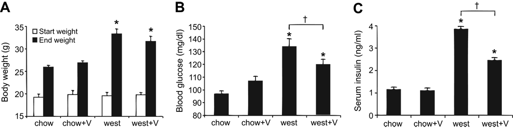
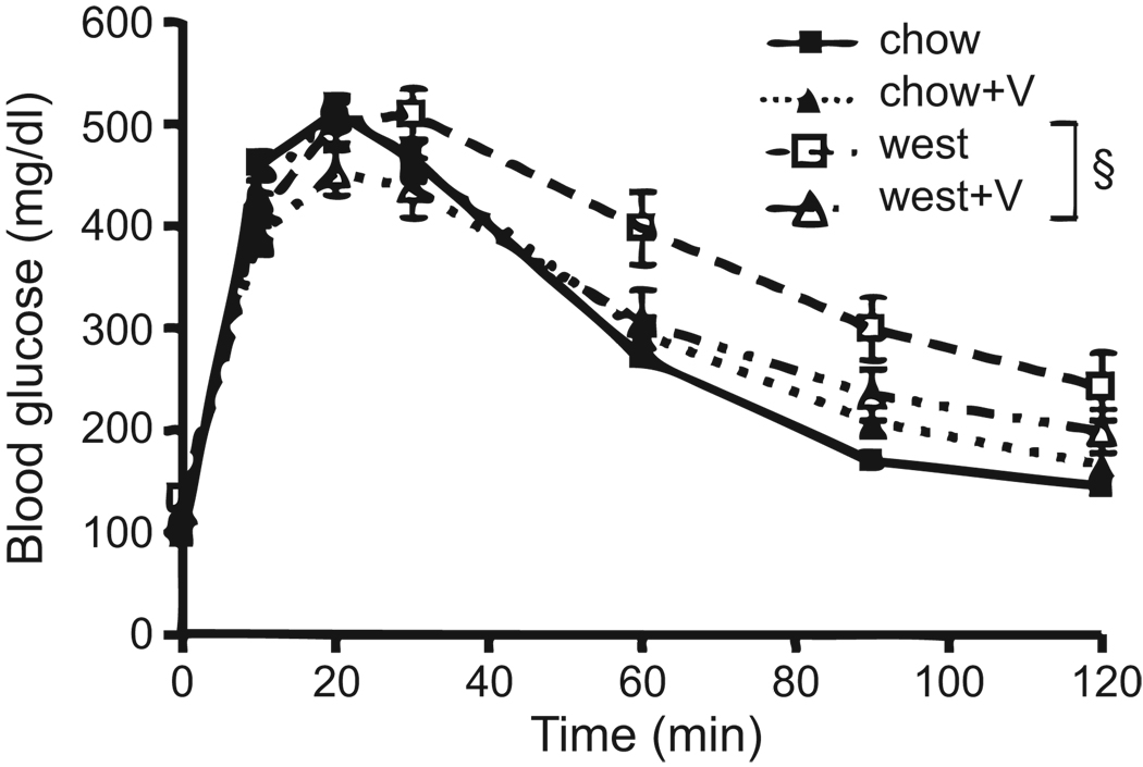
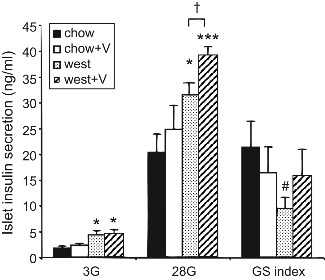
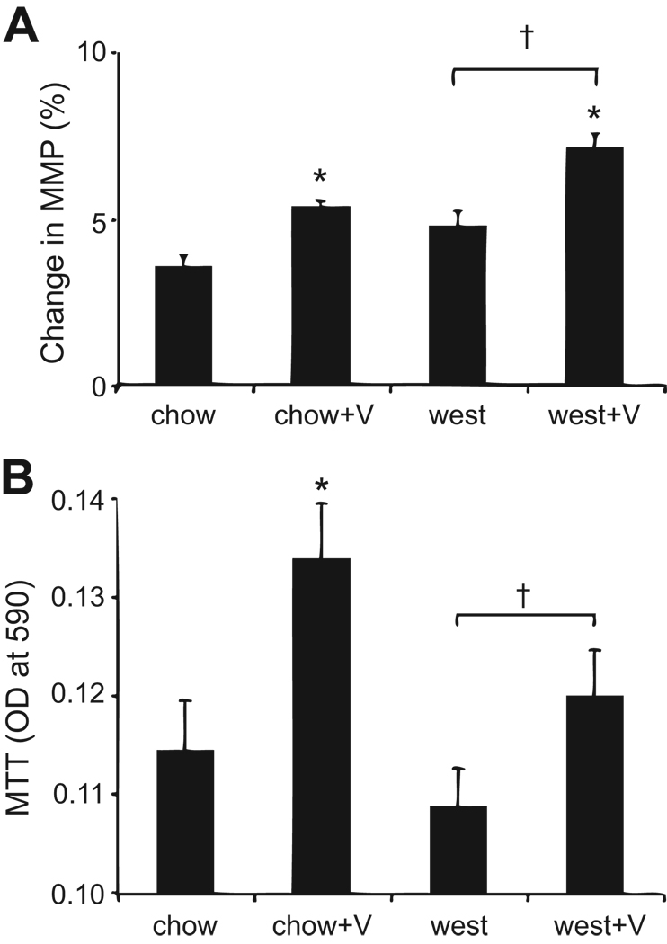
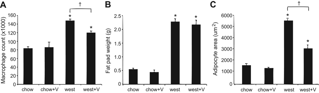
Obesity, hypertension, cardiovascular disease, and inflammation are closely associated with the rising incidence of diabetes mellitus. One pharmacological target that may have significant potential to lower the risk of obesity-related diseases is the angiotensin type 1 receptor (AT1R). We examined the hypothesis that the AT1R blocker valsartan reduces the metabolic consequences and inflammatory effects of a high-fat (Western) diet in mice. C57BL/6J mice were treated by oral gavage with 10 mg/kg per day of valsartan or vehicle and placed on either a standard chow or Western diet for 12 weeks. Western diet-fed mice given valsartan had improved glucose tolerance, reduced fasting blood glucose levels, and reduced serum insulin levels compared with mice fed a Western diet alone. Valsartan treatment also blocked Western diet-induced increases in serum levels of the proinflammatory cytokines interferon-gamma and monocyte chemotactic protein 1. In the pancreatic islets, valsartan enhanced mitochondrial function and prevented Western diet-induced decreases in glucose-stimulated insulin secretion. In adipose tissue, valsartan reduced Western diet-induced macrophage infiltration and expression of macrophage-derived monocyte chemotactic protein 1. In isolated adipocytes, valsartan treatment blocked or attenuated Western diet-induced changes in expression of several key inflammatory signals: interleukin 12p40, interleukin 12p35, tumor necrosis factor-alpha, interferon-gamma, adiponectin, platelet 12-lipoxygenase, collagen 6, inducible NO synthase, and AT1R. Our findings indicate that AT1R blockade with valsartan attenuated several deleterious effects of the Western diet at the systemic and local levels in islets and adipose tissue. This study suggests that AT1R blockers provide additional therapeutic benefits in the metabolic syndrome and other obesity-related disorders beyond lowering blood pressure.
Figures
Comment in
-
Role of renin-angiotensin system blockades in reciprocal relationship between insulin resistance and endothelial dysfunction.Hypertension. 2010 Dec;56(6):e169; author reply e170. doi: 10.1161/HYPERTENSIONAHA.110.161869. Epub 2010 Nov 8. Hypertension. 2010. PMID: 21059993 No abstract available.
Similar articles
-
12-Lipoxygenase-knockout mice are resistant to inflammatory effects of obesity induced by Western diet.Am J Physiol Endocrinol Metab. 2008 Nov;295(5):E1065-75. doi: 10.1152/ajpendo.90371.2008. Epub 2008 Sep 9. Am J Physiol Endocrinol Metab. 2008. PMID: 18780776 Free PMC article.
-
Valsartan improves adipose tissue function in humans with impaired glucose metabolism: a randomized placebo-controlled double-blind trial.PLoS One. 2012;7(6):e39930. doi: 10.1371/journal.pone.0039930. Epub 2012 Jun 29. PLoS One. 2012. PMID: 22768174 Free PMC article. Clinical Trial.
-
Effects of combination therapy with vildagliptin and valsartan in a mouse model of type 2 diabetes.Cardiovasc Diabetol. 2013 Nov 4;12:160. doi: 10.1186/1475-2840-12-160. Cardiovasc Diabetol. 2013. PMID: 24188631 Free PMC article.
-
Adipose inflammation, insulin resistance, and cardiovascular disease.JPEN J Parenter Enteral Nutr. 2008 Nov-Dec;32(6):638-44. doi: 10.1177/0148607108325251. JPEN J Parenter Enteral Nutr. 2008. PMID: 18974244 Free PMC article. Review.
-
Inflammasomes: novel therapeutic targets for metabolic syndrome?Front Endocrinol (Lausanne). 2025 May 13;16:1569579. doi: 10.3389/fendo.2025.1569579. eCollection 2025. Front Endocrinol (Lausanne). 2025. PMID: 40433411 Free PMC article. Review.
Cited by
-
Angiotensin II type 1 receptor antagonist attenuates lacrimal gland, lung, and liver fibrosis in a murine model of chronic graft-versus-host disease.PLoS One. 2013 Jun 6;8(6):e64724. doi: 10.1371/journal.pone.0064724. Print 2013. PLoS One. 2013. PMID: 23762250 Free PMC article.
-
The renin angiotensin aldosterone system and insulin resistance in humans.Curr Hypertens Rep. 2013 Feb;15(1):59-70. doi: 10.1007/s11906-012-0323-2. Curr Hypertens Rep. 2013. PMID: 23242734 Free PMC article. Review.
-
Dipeptidyl peptidase IV inhibitor sitagliptin reduces local inflammation in adipose tissue and in pancreatic islets of obese mice.Am J Physiol Endocrinol Metab. 2011 Feb;300(2):E410-21. doi: 10.1152/ajpendo.00463.2010. Epub 2010 Nov 16. Am J Physiol Endocrinol Metab. 2011. PMID: 21081706 Free PMC article.
-
Chronic AT1 blockade improves glucose homeostasis in obese OLETF rats.J Endocrinol. 2018 Jun;237(3):271-284. doi: 10.1530/JOE-17-0678. Epub 2018 Apr 11. J Endocrinol. 2018. PMID: 29643115 Free PMC article.
-
Benefits of Valsartan and Amlodipine in Lipolysis through PU.1 Inhibition in Fructose-Induced Adiposity.Nutrients. 2022 Sep 12;14(18):3759. doi: 10.3390/nu14183759. Nutrients. 2022. PMID: 36145135 Free PMC article.
References
-
- Olivares-Reyes JA, Arellano-Plancarte A, Castillo-Hernandez JR. Angiotensin II and the development of insulin resistance: Implications for diabetes. Mol Cell Endocrinol. 2009;302:128–139. - PubMed
-
- Abuissa H, Jones PG, Marso SP, O'Keefe JH., Jr Angiotensin-converting enzyme inhibitors or angiotensin receptor blockers for prevention of type 2 diabetes: a meta-analysis of randomized clinical trials. J Am Coll Cardiol. 2005;46:821–826. - PubMed
-
- Mori Y, Itoh Y, Tajima N. Angiotensin II receptor blockers downsize adipocytes in spontaneously type 2 diabetic rats with visceral fat obesity. Am J Hypertens. 2007;20:431–436. - PubMed
-
- Shimabukuro M, Tanaka H, Shimabukuro T. Effects of telmisartan on fat distribution in individuals with the metabolic syndrome. J Hypertens. 2007;25:841–848. - PubMed
-
- Janke J, Engeli S, Gorzelniak K, Luft FC, Sharma AM. Mature adipocytes inhibit in vitro differentiation of human preadipocytes via angiotensin type 1 receptors. Diabetes. 2002;51:1699–1707. - PubMed
Publication types
MeSH terms
Substances
Grants and funding
LinkOut - more resources
Full Text Sources
Other Literature Sources
Medical
Research Materials
