. 2010 Feb;120(2):422-32.
doi: 10.1172/JCI38136. Epub 2010 Jan 25.

TGF-beta activity protects against inflammatory aortic aneurysm progression and complications in angiotensin II-infused mice

Affiliations

TGF-beta activity protects against inflammatory aortic aneurysm progression and complications in angiotensin II-infused mice

Yu Wang et al. J Clin Invest. 2010 Feb.

Abstract

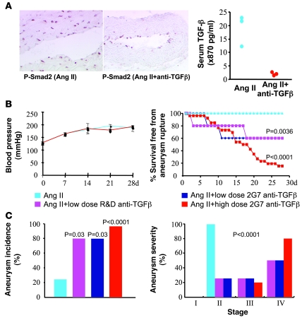
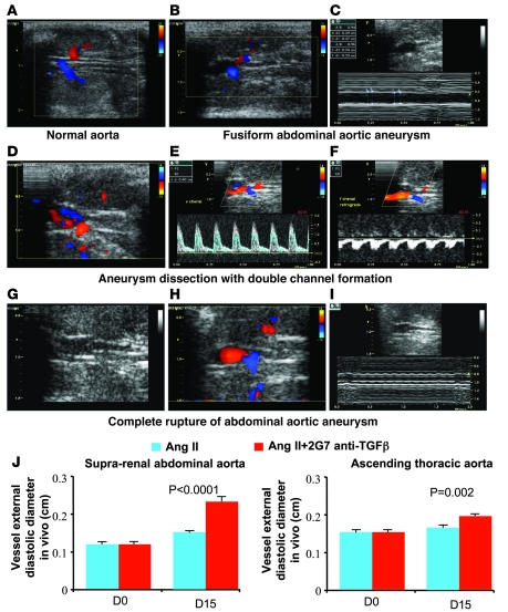
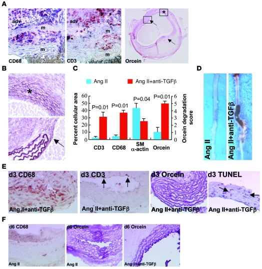
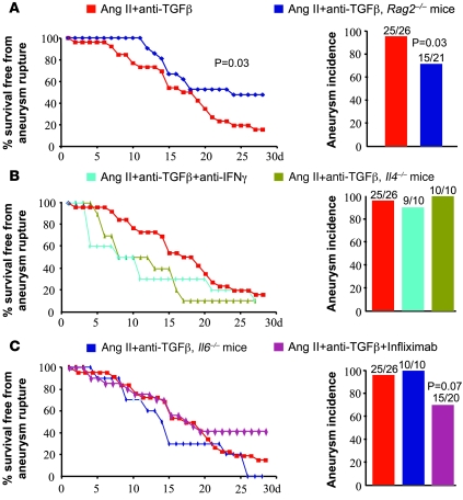
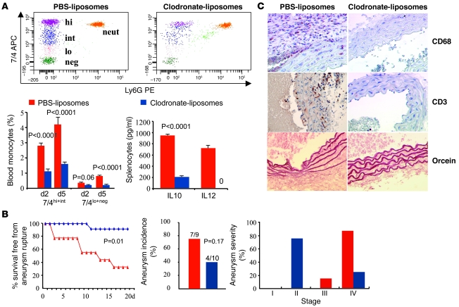
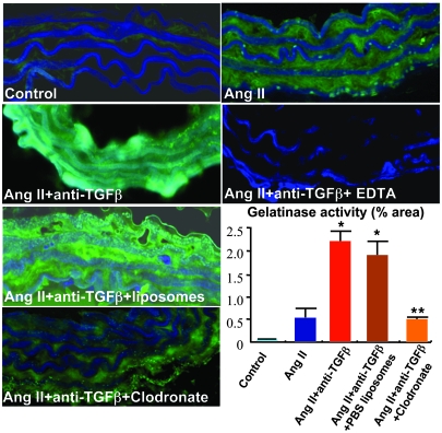
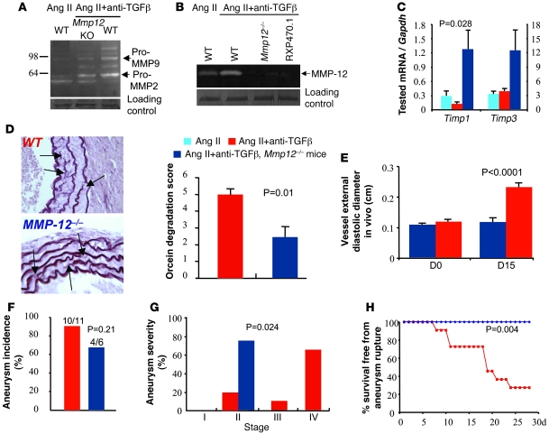
Complicated abdominal aortic aneurysm (AAA) is a major cause of mortality in elderly men. Ang II-dependent TGF-beta activity promotes aortic aneurysm progression in experimental Marfan syndrome. However, the role of TGF-beta in experimental models of AAA has not been comprehensively assessed. Here, we show that systemic neutralization of TGF-beta activity breaks the resistance of normocholesterolemic C57BL/6 mice to Ang II-induced AAA formation and markedly increases their susceptibility to the disease. These aneurysms displayed a large spectrum of complications on echography, including fissuration, double channel formation, and rupture, leading to death from aneurysm complications. The disease was refractory to inhibition of IFN-gamma, IL-4, IL-6, or TNF-alpha signaling. Genetic deletion of T and B cells or inhibition of the CX3CR1 pathway resulted in partial protection. Interestingly, neutralization of TGF-beta activity enhanced monocyte invasiveness, and monocyte depletion markedly inhibited aneurysm progression and complications. Finally, TGF-beta neutralization increased MMP-12 activity, and MMP-12 deficiency prevented aneurysm rupture. These results clearly identify a critical role for TGF-beta in the taming of the innate immune response and the preservation of vessel integrity in C57BL/6 mice, which contrasts with its reported pathogenic role in Marfan syndrome.


Figures

Figure 1
Figure 1. Neutralization of TGF-β activity induces a highly permissive state for Ang II–induced aortic aneurysm formation and complications.
(A) Phosphorylation of Smad-2 (nuclear red staining) is abrogated in the vascular media and adventitia of aortas recovered 3 days after intraperitoneal administration of 500 μg of anti–TGF-β antibody (2G7 clone). Note that at this time point, the media and adventitia are infiltrated by macrophages (see Figure 3E). Original magnification, ×200 for Ang II and ×100 for Ang II + anti–TGF-β. Right panel: Serum TGF-β is barely detectable in mice 3 days after 500 μg of anti–TGF-β antibody compared with nontreated animals. Results are representative of 2 different experiments. (B) Neutralization of TGF-β activity did not alter blood pressure levels (left panel). Right panel: Effects of low-dose anti–TGF-β antibody (R&D Systems, n = 5; or 2G7 clone, n = 5) or high-dose 2G7 clone (n = 26) on survival from ruptured aortic aneurysm, compared with mice that received Ang II in the absence of TGF-β neutralization (n = 29). (C) Quantification of aneurysm incidence and severity (increasing severity from stage I to stage IV as described in ref. 52) in all groups of mice. Stage IV was attributed to ruptured aneurysms.
Figure 2
Figure 2. Two-dimensional color-coded ultrasound imaging of aortic aneurysm development and complications in mice.
(A) Two-dimensional (2D) ultrasound imaging of a normal abdominal aorta visualized between the left (top) and the right (bottom) kidneys. (B) 2D color-coded Doppler imaging revealed a fusiform dilation of the upper renal abdominal aorta. M-mode showed increased diastolic and systolic diameters (C). (DF) Dissected aneurysm of the upper renal abdominal aorta. Color-coded Doppler imaging revealed a dissection with 2 lumens: the true lumen was coded in red and showed a forward blood flow toward the renal arteries and the suprarenal abdominal aorta; the second lumen, colored in blue, showed a backward blood flow (D). Pulsed Doppler revealed an accelerated forward blood flow in the true lumen (E) and a backward blood flow in the second lumen (F), which was suggestive of false channel formation. (GI) Complete disintegration of the distal part of an aortic aneurysm (G). 2D color-coded Doppler imaging evidenced the inferior limit of the aneurysm just upstream of the emergence of the right renal artery coded in blue (H). Targeted M-mode is shown in I. (J) Histograms show determination of vessel diameter in vivo using ultrasound imaging at day 0 and day 15 after the beginning of treatment. Vessel dilatation at the suprarenal level was markedly pronounced in the mice that received anti–TGF-β antibody (n = 7 for Ang II and n = 17 for Ang II + anti-TGF-β). We also found a significant dilatation of the ascending aorta at day 15 in the anti–TGF-β group (n = 7–9 per group).
Figure 3
Figure 3. Characterization of Ang II–induced aortic aneurysms in mice treated with anti–TGF-β antibody.
(A) Representative photomicrographs of macrophage (CD68), T lymphocyte (CD3), and elastin (orcein) stainings of aortic aneurysms from mice infused with Ang II and treated with anti–TGF-β antibody. adv, adventitia; m, media. Arrows show the sites of complete elastin degradation. (B) Higher magnifications of vessel areas indicated in the right panel of A. Asterisk indicates advanced elastin disruption of all lamellae. Arrow shows the edge of degraded elastin lamellae. (C) Quantification (n = 5 mice per group) of smooth muscle cell (α-actin) content, elastin (orcein) degradation, as well as macrophage and T lymphocyte infiltration. (D) Representative photomicrographs of aortas recovered from mice infused with Ang II in the presence or absence of anti–TGF-β antibody. (E) Aortic tissue sections recovered after 3 days of treatment and stained for the presence of macrophages (CD68, brown), T lymphocytes (CD3, punctuate red-brown, arrows), elastin (orcein), or apoptotic cells (TUNEL, red-brown nuclei, arrows). Note that macrophage infiltration preceded T cell accumulation and overt elastin degradation and coincided with the occurrence of VSMC death. (F) Staining for macrophages (CD68) and elastin (orcein) after 6 days of treatment. Rare macrophages and no elastin degradation were observed in the Ang II group. However, intense elastin degradation (2.4 ± 0.4 elastin lamellae lost) was detected at this time point in the Ang II + anti–TGF-β group. Data are representative of 5 mice per time point and per group. Original magnification, ×400 (A, except right panel: ×40); ×400 (B); ×100 (E, except right panel: ×200); ×200 (F).
Figure 4
Figure 4. Ang II–induced aortic aneurysms in mice treated with anti–TGF-β antibody are not prevented by inhibition of IFN-γ, IL-4, IL-6, or TNF-α signaling.
Kaplan-Meier curves of survival free from aneurysm rupture and quantification of aortic aneurysm incidence in all groups of mice. (A) Deficiency of T and B cells in Rag2–/– mice (n = 21) slightly but significantly reduced aneurysm incidence and severity compared with controls (n = 26). (B and C) However, inhibition of IFN-γ (n = 10) or TNF-α (n = 20) signaling or genetic deficiency in IL-4 (n = 10) or IL-6 (n = 10) did not afford protection. Results for the Ang II + anti–TGF-β group are shown only for comparison, but the experiments were not conducted simultaneously with the other groups of mice.
Figure 5
Figure 5. Ang II–induced aortic aneurysm in mice treated with anti–TGF-β antibody is prevented by monocyte depletion.
(A) Representative examples of flow cytometry and quantitative analysis of the various monocyte subsets in mice treated with either PBS- or clodronate-containing liposomes. Clodronate treatment significantly reduced the number of all monocyte subsets, with no significant effects on neutrophil count. Results are mean ± SEM of 6 mice per group. Clodronate treatment also reduced IL-10 and abrogated IL-12 production by LPS/INF-γ–treated splenocytes recovered from clodronate-treated animals (results are representative of 3 mice per group). (B) Kaplan-Meier curves of survival free from aneurysm rupture and quantification of aortic aneurysm incidence and severity in mice (Ang II + anti–TGF-β) treated with either PBS- (n = 9) or clodronate-liposomes (n = 10). P = 0.026, clodronate- versus PBS-liposomes. (C) Representative photomicrographs of macrophage (CD68) and T lymphocyte (CD3) infiltration as well as elastin content (orcein) of the aortic vessel wall in mice treated with either PBS- or clodronate-liposomes. Original magnification, ×400.
Figure 6
Figure 6. Neutralization of TGF-β induces a monocyte/macrophage-dependent increase in gelatinase activity in the abdominal aorta.
Representative examples and semiquantitative analysis (percent positivity relative to the whole section area) of gelatinase activity (green) in suprarenal abdominal aortas of control mice; mice infused with Ang II, either with or without anti–TGF-β; and those treated with anti–TGF-β and either PBS- or clodronate-containing liposomes. Elastin lamellae are depicted in blue. TGF-β neutralization led to a marked increase in gelatinase activity (typical areas with enhanced gelatinolytic activity are shown), which was significantly inhibited by depletion of monocytes/macrophages using clodronate. EDTA in vitro completely abrogated gelatinase activity. Results are mean ± SEM of 4–5 mice per group. Original magnification, ×400. *P < 0.05, **P < 0.01.
Figure 7
Figure 7. MMP-12 is required for aneurysm progression and rupture in mice treated with Ang II and anti–TGF-β antibody.
Representative examples of 5 separate experiments showing gelatinolytic MMP-9 and MMP-2 (A) and caseinolytic MMP-12 (B) activities in suprarenal abdominal aortas of mice infused with Ang II, with or without anti-TGF-β, and with or without genetic deficiency for Mmp12. RXP470.1 completely inhibited the caseinolytic activity, further indicating that the activity was specific for MMP-12. Loading controls represent bands obtained after Coomassie blue staining. For technical reasons, loading control of the casein zymography was run on a separate gel. (C) Quantification of aortic mRNA expression (Q-PCR) of Timp1 and Timp3 (n = 5–6 per group). We found a significant increase in Timp1 expression in Mmp12-deficient mice. (D) Representative examples and quantitative analysis of elastin (orcein staining) degradation (arrows) in WT and Mmp12-deficient mice infused with Ang II and treated with anti–TGF-β antibody. Data were collected using aortic specimens recovered at day 28 or at necropsy when animal death from AAA rupture occurred before day 28. Original magnification, ×400. (E) Vessel diameter in vivo using ultrasound imaging. Targeted M-mode–derived blinded measurements of aortic diameters were obtained as described in Methods at days 0 and 15 after the beginning of treatment. Vessel dilatation at the suprarenal level was abrogated at day 15 in Mmp12-deficient mice. (FH) Quantification of aortic aneurysm incidence (F) and severity (G) as well as Kaplan-Meier curves of survival free from aneurysm rupture (H) in WT and Mmp12–deficient mice. Mmp12 deficiency significantly reduced aneurysm severity and completely protected from aneurysm rupture.

Comment in

Similar articles

Cited by

References

    1. Best VA, Price JF, Fowkes FG. Persistent increase in the incidence of abdominal aortic aneurysm in Scotland, 1981-2000. Br J Surg. 2003;90(12):1510–1515. - PubMed
    1. Fleming C, Whitlock EP, Beil TL, Lederle FA. Screening for abdominal aortic aneurysm: a best-evidence systematic review for the U.S. Preventive Services Task Force. Ann Intern Med. 2005;142(3):203–211. - PubMed
    1. Golledge J, Muller J, Daugherty A, Norman P. Abdominal aortic aneurysm: pathogenesis and implications for management. Arterioscler Thromb Vasc Biol. 2006;26(12):2605–2613. doi: 10.1161/01.ATV.0000245819.32762.cb. - DOI - PubMed
    1. Powell JT, Brady AR. Detection, management, and prospects for the medical treatment of small abdominal aortic aneurysms. Arterioscler Thromb Vasc Biol. 2004;24(2):241–245. doi: 10.1161/01.ATV.0000106016.13624.4a. - DOI - PubMed
    1. Golledge J, et al. Association between osteopontin and human abdominal aortic aneurysm. Arterioscler Thromb Vasc Biol. 2007;27(3):655–660. doi: 10.1161/01.ATV.0000255560.49503.4e. - DOI - PubMed

Publication types

MeSH terms

Substances