Bone marrow cell cotransplantation with islets improves their vascularization and function
- PMID: 20101199
- PMCID: PMC2844476
- DOI: 10.1097/TP.0b013e3181cb3e8d
Bone marrow cell cotransplantation with islets improves their vascularization and function
Abstract
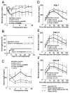
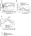
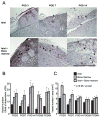
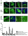
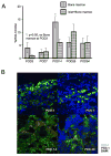
BACKGROUND.: To test the angiogenesis-promoting effects of bone marrow cells when cotransplanted with islets. METHODS.: Streptozotocin-induced diabetic BALB/c mice were transplanted syngeneically under the kidney capsule: (1) 200 islets, (2) 1 to 5x10 bone marrow cells, or (3) 200 islets and 1 to 5x10 bone marrow cells. All mice were evaluated for blood glucose, serum insulin, and glucose tolerance up to postoperative day (POD) 28, and a subset was monitored for 3 months after transplantation. Histologic assessment was performed at PODs 3, 7, 14, 28, and 84 for the detection of von Willebrand factor (vWF), vascular endothelial growth factor (VEGF), insulin, cluster of differentiation-34, and pancreatic duodenal homeobox-1 (PDX-1) protein. RESULTS.: Blood glucose was the lowest and serum insulin was the highest in the islet+bone marrow group at POD 7. Blood glucose was significantly lower in the islet+bone marrow group relative to the islet only group after 63 days of transplantation (P<0.05). Significantly more new periislet vessels were detected in the islet+bone marrow group compared with the islet group (P<0.05). Vascular endothelial growth factor staining was more prominent in bone marrow than in islets (P<0.05). Pancreatic duodenal homeobox-1-positive areas were identified in bone marrow cells with an increase in staining over time. However, there were no normoglycemic mice and no insulin-positive cells in the bone marrow alone group. CONCLUSIONS.: Cotransplantation of bone marrow cells with islets is associated with enhanced islet graft vascularization and function.
Figures

Similar articles
-
Bone marrow cells produce nerve growth factor and promote angiogenesis around transplanted islets.World J Gastroenterol. 2010 Mar 14;16(10):1215-20. doi: 10.3748/wjg.v16.i10.1215. World J Gastroenterol. 2010. PMID: 20222164 Free PMC article.
-
Nerve growth factor is associated with islet graft failure following intraportal transplantation.Islets. 2012 Jan-Feb;4(1):24-31. doi: 10.4161/isl.18467. Epub 2011 Dec 23. Islets. 2012. PMID: 22192949 Free PMC article.
-
Skeletal Myoblast Cells Enhance the Function of Transplanted Islets in Diabetic Mice.J Diabetes Res. 2024 May 17;2024:5574968. doi: 10.1155/2024/5574968. eCollection 2024. J Diabetes Res. 2024. PMID: 38800586 Free PMC article.
-
Gene expression and silencing for improved islet transplantation.J Control Release. 2009 Dec 16;140(3):262-7. doi: 10.1016/j.jconrel.2009.04.011. Epub 2009 Apr 17. J Control Release. 2009. PMID: 19376168 Free PMC article. Review.
-
Roles for bone-marrow-derived cells in beta-cell maintenance.Trends Mol Med. 2004 Nov;10(11):558-64. doi: 10.1016/j.molmed.2004.09.002. Trends Mol Med. 2004. PMID: 15519282 Review.
Cited by
-
Mesenchymal stem cells in the treatment of type 1 diabetes mellitus.Endocr Pathol. 2015 May;26(2):95-103. doi: 10.1007/s12022-015-9362-y. Endocr Pathol. 2015. PMID: 25762503 Review.
-
Utility of co-transplanting mesenchymal stem cells in islet transplantation.World J Gastroenterol. 2011 Dec 21;17(47):5150-5. doi: 10.3748/wjg.v17.i47.5150. World J Gastroenterol. 2011. PMID: 22215938 Free PMC article. Review.
-
Extracellular Matrix and Growth Factors Improve the Efficacy of Intramuscular Islet Transplantation.PLoS One. 2015 Oct 16;10(10):e0140910. doi: 10.1371/journal.pone.0140910. eCollection 2015. PLoS One. 2015. PMID: 26473955 Free PMC article.
-
The effect of diabetes mellitus on differentiation of mesenchymal stem cells into insulin-producing cells.Biol Res. 2024 May 2;57(1):20. doi: 10.1186/s40659-024-00502-4. Biol Res. 2024. PMID: 38698488 Free PMC article.
-
Mesenchymal stem cells as feeder cells for pancreatic islet transplants.Rev Diabet Stud. 2010 Summer;7(2):132-43. doi: 10.1900/RDS.2010.7.132. Epub 2010 Aug 10. Rev Diabet Stud. 2010. PMID: 21060972 Free PMC article. Review.
References
-
- Ryan EA, Paty BW, Senior PA, et al. Five-year follow-up after clinical islet transplantation. Diabetes. 2005;54(7):2060. - PubMed
-
- Shapiro AM, Nanji SA, Lakey JR. Clinical islet transplant: current and future directions towards tolerance. Immunol Rev. 2003;196:219. - PubMed
-
- Shapiro AM, Lakey JR, Ryan EA, et al. Islet transplantation in seven patients with type 1 diabetes mellitus using a glucocorticoid-free immunosuppressive regimen. N Engl J Med. 2000;343(4):230. - PubMed
-
- Stagner JI, Mokshagundam S, Samols E. Hormone secretion from transplanted islets is dependent upon changes in islet revascularization and islet architecture. Transplant Proc. 1995;27(6):3251. - PubMed
-
- Emamaullee JA, Rajotte RV, Liston P, et al. XIAP overexpression in human islets prevents early posttransplant apoptosis and reduces the islet mass needed to treat diabetes. Diabetes. 2005;54(9):2541. - PubMed
MeSH terms
Substances
Grants and funding
LinkOut - more resources
Full Text Sources
Other Literature Sources
Medical
Research Materials
Miscellaneous

