. 2010 Apr 22;29(16):2404-14.
doi: 10.1038/onc.2009.520. Epub 2010 Jan 25.

Phosphorylation of the mutant K303R estrogen receptor alpha at serine 305 affects aromatase inhibitor sensitivity

Affiliations

Phosphorylation of the mutant K303R estrogen receptor alpha at serine 305 affects aromatase inhibitor sensitivity

I Barone et al. Oncogene. .

Abstract

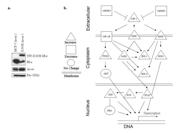
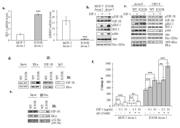
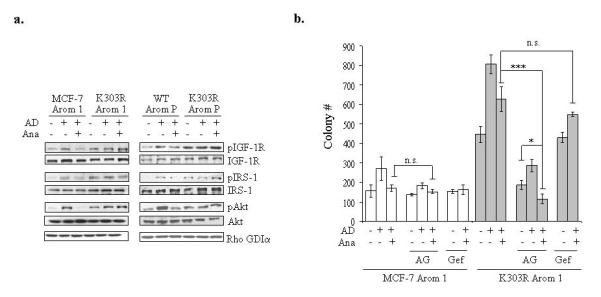
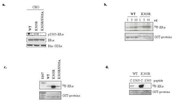
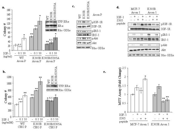
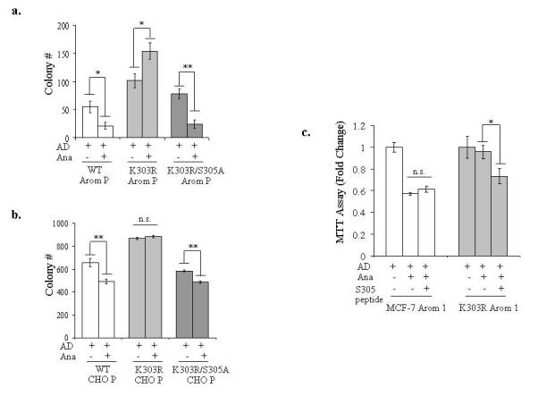
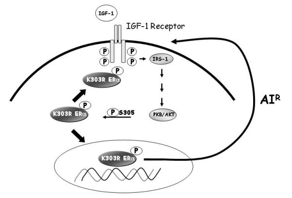
We earlier identified a lysine to arginine transition at residue 303 (K303R) in estrogen receptor alpha (ERalpha) in invasive breast cancers, which confers resistance to the aromatase inhibitor (AI) anastrozole (Ana) when expressed in MCF-7 breast cancer cells. Here, we show that AI resistance arises through an enhanced cross talk of the insulin-like growth factor receptor-1 (IGF-1R)/insulin receptor substrate (IRS)-1/Akt pathway with ERalpha, and the serine (S) residue 305 adjacent to the K303R mutation has a key function in mediating this cross talk. The ERalpha S305 residue is an important site that modifies response to tamoxifen; thus, we questioned whether this site could also influence AI response. We generated stable transfectants-expressing wild-type, K303R ERalpha or a double K303R/S305A mutant receptor, and found that the AI-resistant phenotype associated with expression of the K303R mutation was dependent on activation of S305 within the receptor. Ana significantly reduced growth in K303R/S305A-expressing cells. Preventing S305 phosphorylation with a blocking peptide inhibited IGF-1R/IRS-1/Akt activation and also restored AI sensitivity. Our data suggest that the K303R mutation and the S305 ERalpha residue may be a novel determinant of AI response in breast cancer, and blockade of S305 phosphorylation represents a new therapeutic strategy for treating tumors resistant to hormone therapy.


Figures

Figure 1
Figure 1
(a) Immunoblot analysis for ERα and aromatase (Arom) expression in MCF-7 Arom 1 and K303R Arom 1-expressing cells. Rho GDIα was used as a control for equal loading and transfer. (b) Schematic representation of gene expression changes of insulin/IGF family members of K303R-expressing cells (n=4) compared to WT cells (n=4) studied by microarray analysis. The genes that were induced are represented in triangles; those that were repressed are indicated with a square, and unchanged genes are shown with a circle.
Figure 2
Figure 2
The K303R ERα mutant cells exhibited increased activation of the IGF-1 signaling pathway. (a) IRS-1 and IGFBP3 mRNA expression was assessed by quantitative real-time RT-PCR analysis. Each bar represents mRNA expression relative to β-actin mRNA (endogenous control gene) ± SD (average of four samples). ***P<0.0001 K303R-Arom 1 mRNA levels compared to MCF-7 Arom 1 (b, c) Serum-deprived MCF-7 Arom 1 and K303R Arom 1 cells (b) or MCF-7 WT, K303R P and CHO WT, K303R P (c) pools of stably transfected and overexpressing cells were treated with vehicle or IGF-1 10ng/ml for 5min. Total cellular extracts were analyzed for phosphorylation (p) and expression of IGF-1R, IRS-1 and Akt by immunoblot analysis. Rho GDIα was used as a control for equal loading and transfer. Immunoblots show a single representative of three separate experiments. (d, e) Lysates from CHO cells transiently transfected with WT or K303R ERα were immunoprecipitated (IP) with anti-ERα or IGF-1R or IgG (negative control) (d) or Shc (e) antibodies and immunoblotted for IGF-1R, ERα or Shc. (f) Cells were plated in soft agar, and then treated with vehicle (−) or increasing amounts of IGF-1 (0.1 and 10ng/ml) ± AD 10nM. Cells were allowed to grow for 14 days, and the number of colonies >50μm were quantified, and the results were graphed. Data are the mean colony number of three plates and representative of two independent experiments. Bars, SD. n.s.=nonsignificant, **P<0.01, ***P<0.0005.
Figure 3
Figure 3
Inhibition of the IGF-1R pathway reversed AIR. (a) MCF-7 Arom 1 and K303R Arom 1 cells or MCF-7 WT, K303R P pools of stably transfected and overexpressing cells (WT Arom P and K303R Arom P) were treated with vehicle (C), AD 10nM ± anastrozole (Ana 1μM) for 1h. Levels of phosphorylated (p) IGF-1R, IRS-1 and Akt and total non-phosphorylated proteins were measured using immunoblot analysis. Rho GDIα was used as a control for equal loading and transfer. Immunoblots shown are a single representative blot of two separate experiments. (b) Cells were plated in soft agar and then treated with vehicle, AD 10nM, Ana 1μM with or without the IGF-1R inhibitor AG1024 (AG 10μM) or the EGFR inhibitor Gefitinib (Gef, 1μM). The number of colonies >50μm were quantified, and data are the mean colony number of three plates and representative of three independent experiments. Bars, SD. n.s.=nonsignificant, *P=0.02, ***P<0.0005.
Figure 4
Figure 4
The S305 residue of the K303R mutant receptor is a novel Akt phosphorylation site. (a) Lysates from CHO cells transiently transfected with WT, K303R ERα or K303R/S305A ERα plasmids were analyzed for phosphorylation levels of S305 YFP ERα (pS305) and total non-phosphorylated YFP- ERα using immunoblot analysis. Rho GDIα was used as a control for equal loading and transfer. (b) In vitro Akt kinase assays using increasing concentrations (1, 5, 10μg) of WT and K303R ERα GST-fusion hinge fragments (residues 253-310) were performed in kinase buffer. Reactions products were analyzed using SDS-PAGE, and phosphorylated ERα bands were visualized by autoradiography. (c) In vitro Akt kinase assays using WT, K303R and K303R/S305A ERα GST-fusion fragments. The ERα fragment containing the known S167 site was used as positive control in the reaction. (d) In vitro Akt kinase assays using WT, K303R ERα GST-fusion fragments were performed in the presence of the S305 blocking or a control (C) peptide. The input GST proteins were visualized with Ponceau S staining (bottom panels).
Figure 5
Figure 5
Inhibition of serine 305 ERα phosphorylation blocked IGF-1R and mutant ERα cross-talk. (a, b) MCF-7 WT, K303R and K303R/S305A Arom P (a) and CHO WT, K303R and K303R/S305A P (b) pools of stably transfected and overexpressing cells were plated in soft agar and then treated with vehicle, IGF-1 0.1 or 10ng/ml for 14 days. The number of colonies >50μm were quantified and data are the mean colony number of three plates and representative of two independent experiments. Bars, SD. n.s.=nonsignificant, *P<0.01, **P<0.005. Equal expression of protein was determined by immunoblot with anti-ERα and Rho GDIα antibodies (right panel for a and b). (c) Cellular extracts from serum-deprived MCF-7 Arom WT, K303R K303R/S305A P pools of stably transfected and overexpressing cells were analyzed for phosphorylation (p) and expression of IGF-1R, IRS-1 and Akt by immunoblot analysis. Rho GDIα was used as a control for equal loading and transfer. (d) Cells were incubated with the S305 peptide (4μg/well) for 4h, and then treated with or without IGF-1 (10ng/ml) for 5min. Levels of phosphorylated (p) IGF-1R, IRS-1 and Akt, and total non-phosphorylated proteins were measured in cellular extracts by immunoblot analysis. Blots are representative of three separate experiments. Rho GDIα was used as a control for equal loading and transfer. (e) MTT growth assays in MCF-7 Arom 1 and K303R Arom 1 cells treated for 3 days with vehicle, IGF-1 (10ng/ml) and/or the S305 peptide (0.6μg/well). Cell proliferation is expressed as fold change relative to vehicle-treated cells. The data are representative of four independent experiments, each performed in triplicate. Columns, mean. Bars, SD. *P<0.005 compared to IGF-1 treatment in MCF-7 Arom 1-cells, **P<0.001 compared to vehicle and IGF-1 treatments in K303R Arom1-expressing cells.
Figure 6
Figure 6
The S305 residue in the K303R ERα mutant is a determinant of AIR. (a, b) MCF-7 WT, K303R and K303R S305A Arom P (a) and CHO WT, K303R and K303R S305A P (b) pools of stably transfected cells were plated in soft agar and then treated with AD (10nM) ± Ana (1μM) for 14 days. The number of colonies >50μm were quantified, and data shown are the mean colony number of two plates and representative of three independent experiments. Bars, SD. n.s.= nonsignificant, *P<0.01, **P<0.005. (c) MTT growth assay in MCF-7 Arom 1 and K303R Arom 1 cells treated for 6 days with AD (10nM), Ana (1μM) and/or the S305 peptide (0.6μg/well). Cell proliferation is expressed as fold change relative to AD-treated cells. The data are representative of three independent experiments, each performed in triplicate. Columns, mean. Bars, SD. n.s.=nonsignificant, *P=0.01 AD+Ana+S305 peptide compared to Ana+AD.
Figure 7
Figure 7
Model depicting potential cross-talk between the ERα and IGF-signaling components in breast cancer cells expressing the mutation. K303R ERα mutant receptor binds to IGF-1R and activates the IGF-1R signaling cascades, including IRS-1 and Akt pathways. Activated Akt promotes the phosphorylation of the K303R ERα mutant at the S305 residue, leading to an increase in the transcription of IGF signaling molecules, thus establishing a positive feedback loop between growth factor and steroid receptors that leads to AIR.

Similar articles

Cited by

References

    1. Agoulnik IU, Tong XW, Fischer DC, Korner K, Atkinson NE, Edwards DP, et al. A germline variation in the progesterone receptor gene increases transcriptional activity and may modify ovarian cancer risk. J Clin Endocrinol Metab. 2004;89:6340–7. - PubMed
    1. Arteaga CL, Osborne CK. Growth inhibition of human breast cancer cells in vitro with an antibody against the type I somatomedin receptor. Cancer Res. 1989;49:6237–41. - PubMed
    1. Backer JM, Myers MG, Jr., Shoelson SE, Chin DJ, Sun XJ, Miralpeix M, et al. Phosphatidylinositol 3′-kinase is activated by association with IRS-1 during insulin stimulation. EMBO J. 1992;11:3469–79. - PMC - PubMed
    1. Barone I, Cui Y, Herynk MH, Corona-Rodriguez A, Giordano C, Selever J, et al. Expression of the K303R estrogen receptor-alpha breast cancer mutation induces resistance to an aromatase inhibitor via addiction to the PI3K/Akt kinase pathway. Cancer Res. 2009;69:4724–32. - PMC - PubMed
    1. Blume-Jensen P, Hunter T. Oncogenic kinase signalling. Nature. 2001;411:355–65. - PubMed

Publication types