. 2010 Apr 8;29(14):2047-59.
doi: 10.1038/onc.2009.486. Epub 2010 Jan 18.

TrkB induces EMT and has a key role in invasion of head and neck squamous cell carcinoma

Affiliations

TrkB induces EMT and has a key role in invasion of head and neck squamous cell carcinoma

M E Kupferman et al. Oncogene. .

Abstract

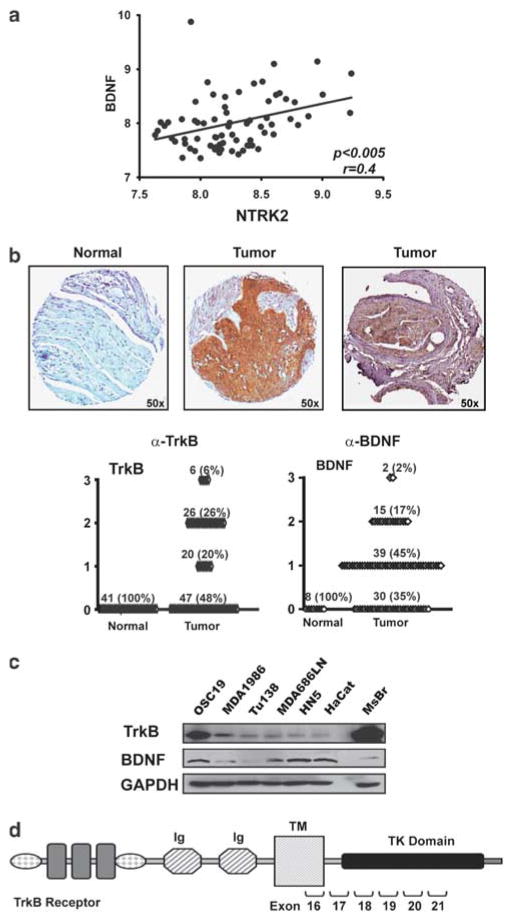
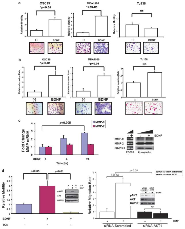
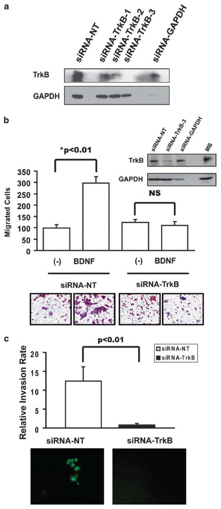
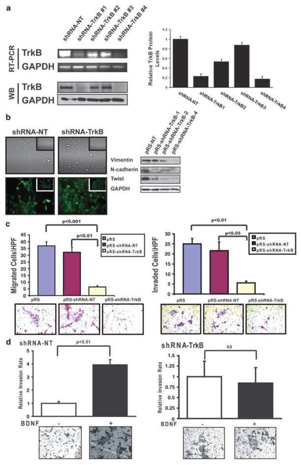
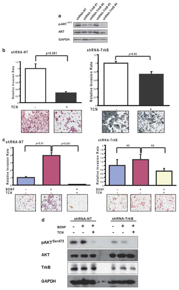
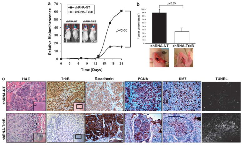
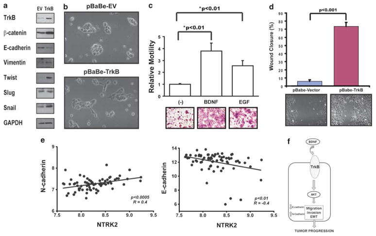
Head and neck squamous cell carcinoma (HNSCC) remains a significant public health problem, accounting for over 5% of all cancer-related deaths, and these deaths primarily result from metastatic disease. The molecular processes involved in HNSCC pathogenesis and progression are poorly understood, and here we present experimental evidence for a direct role of the cell surface receptor tyrosine kinase, TrkB, in HNSCC tumor progression. Using immunohistochemical analysis and transcriptional profiling of archival HNSCC tumor specimens, we found that TrkB and its secreted ligand, brain-derived neurotrophic factor (BDNF), are expresses in greater than 50% of human HNSCC tumors, but not in normal upper aerodigestive tract (UADT) epithelia. Studies with HNSCC cell lines reveal that in vitro stimulation with BDNF, the ligand for TrkB, upregulates the migration and invasion of HNSCC cells, and both transient and stable suppressions of TrkB result in significant abrogation of constitutive and ligand-mediated migration and invasion. Furthermore, enforced overexpression of TrkB results in altered expression of molecular mediators of epithelial-to-mesenchymal transition (EMT), including downregulation of E-cadherin and upregulation of Twist. Using an in vivo mouse model of HNSCC, we were able to show that downregulation of TrkB suppresses tumor growth. These results directly implicate TrkB in EMT and the invasive behavior of HNSCC, and correlate with the in vivo overexpression of TrkB in human HNSCC. Taken together, these data suggest that the TrkB receptor may be a critical component in the multi-step tumor progression of HNSCC, and may be an attractive target for much needed new therapies for this disease.


Conflict of interest statement

Conflict of interest

The authors declare no conflict of interest.

Figures

Figure 1
Figure 1
Tyrosine kinase B (TrkB) is over-expressed in human head and neck squamous cell carcinoma (HNSCC) tumors. (a) Complementary DNA (cDNA) microarray analysis revealed that messenger RNA (mRNA) expression of NTRK2 correlated with expression of BDNF in 71 previously untreated human tumors (P<0.005) (b) Expression of TrkB and brain-derived neurotrophic factor (BDNF) in representative samples on a human HNSCC tissue array. Tumors were analyzed and graded for TrkB (P<0.0001) and BDNF (P<0.001) expression (scale: 0–3). (c) Protein lysates from subconfluent HNSCC or normal cell lines were separated by SDS–PAGE and assessed with the indicated antibodies. Mouse brain (MsBr) was used as a positive control for TrkB expression. (d) Schematic representation of the TrkB receptor. Exon numbers indicate regions of the receptor that were assessed for genomic mutational analysis (see Table 1). TM, transmembrane domain; TK, tyrosine kinase.
Figure 2
Figure 2
Tyrosine kinase B (TrkB) expression is associated with differential chemotactic responsiveness to brain-derived neurotrophic factor (BDNF). (a) Head and neck squamous cell carcinoma (HNSCC) cell lines were exposed to BDNF (100 ng/ml) in Transwell migration plates and assessed for chemotactic cellular migration after 24 h. Cells were counted in five high-powered fields, analyzed for differential migration with ImagePro. Epidermal growth factor (EGF) (100 ng/ml) was added to a separate well as a positive control for migration experiments (data not shown). Columns, relative migration of cells in the presence or absence of BDNF; bars, s.e.m; NS, not significant. Representative experimental results from triplicate repeats. Magnification: ×100. (b) HNSCC cell lines were exposed to BDNF in Matrigel invasion plates and assessed for cellular invasion after 24 h. Cells were counted in five high-powered fields and analyzed for differential migration with ImagePro. EGF was added to a separate well as a positive control for invasion experiments (data not shown). Columns, relative invasion of cells in the presence or absence of BDNF; bars, s.e.m; NS, not significant. Magnification: ×100. (c) BDNF induces matrix metallopeptidase 9 (MMP-9) expression and activation in HNSCC. Quantitative RT–PCR (left) and gelatin zymography (right) were performed to determine the induction of MMP expression and function under the control of BDNF stimulation. (d) Cell lines were exposed to BDNF (100 ng/ml) with or without triciribine (5 μM) in migration plates and assessed for migration after 24 h (left). Cells were counted in five high-powered fields and analyzed for differential migration. In parallel, OSC19 cell lines were exposed to BDNF (100 ng/ml) with or without triciribine (5 μM) in 6-well plates after 24 h of serum starvation (left, inset). Cells were collected, lysed and proteins were separated by 10% SDS–PAGE and analyzed for the indicated antibodies. OSC19 cell lines were exposed to BDNF (100 ng/ml) after transfection with small interfering RNA (siRNA) targeting AKT1 or a scrambled sequence in 6-well plates after 24 h of serum starvation (right). Cells were collected, lysed and proteins were separated by 10% SDS–PAGE and analyzed for indicated antibodies.
Figure 3
Figure 3
Transient manipulation of tyrosine kinase B (TrkB) in head and neck squamous cell carcinoma (HNSCC) alters ligand-mediated migration and invasion. (a) OSC19 cells were transiently transfected with siRNA constructs, and lysates from subconfluent cells were analyzed by SDS–PAGE with the indicated antibodies. Glyceraldehyde-3-phosphate dehydrogenase (GAPDH) knockdown was assayed as a positive control. (b) OSC19 cells were transiently transfected with small interfering RNA (siRNA) against TrkB (construct 3) or a non-targeted siRNA (inset), and seeded on Transwell migration plates for 24 h in the presence or absence of BDNF (100 ng/ml). Cells were counted in five high-powered fields and analyzed for differential migration with ImagePro. Experiments were performed in triplicate and repeated three times. Columns, number of migrated cells in the presence or absence of BDNF; bars, s.e.m.; NS, not significant; magnification: × 100. (c) OSC19-Luc cells were transiently transfected with siRNA against TrkB (construct 3) or a non-targeted siRNA, and seeded on Matrigel invasion plates for 24 h. Cells were counted with an inverted fluorescence microscope in five high-powered fields and analyzed for differential invasion with ImagePro. Experiments were performed in triplicate and repeated three times. Columns, relative invasion of cells; bars, s.e.m, magnification: × 100.
Figure 4
Figure 4
Morphological and functional alterations induced by targeted reduction of tyrosine kinase B (TrkB). (a) Messenger RNA (mRNA) (top) and protein (bottom) levels of TrkB after stable transfection of OSC19-Luc cells with vectors targeting an irrelevant sequence (shRNA-NT) or TrkB (shRNA-TrkB). Quantification with ImagePro was performed to determine the relative change in TrkB protein expression among the various constructs (right). (b) Cellular morphology of stably transfected cells ( ×200, inset ×40) under both light (top) and fluorescence (bottom) microscopy. Deregulation of epithelial-to-mesenchymal transition (EMT) markers in transfected cells was determined by 10% SDS–PAGE and assayed with the indicated antibodies (right). (c) Constitutive migration (left) and invasion (right) of parental (pRS), shRNA-NT or shRNA-TrkB transfected cells toward a 10% fetal bovine serum (FBS) gradient. Experiments were performed in triplicate and repeated three times. Columns, relative migration or invasion of cells; bars, s.e.m.; magnification: ± 100. (d) brain-derived neurotrophic factor (BDNF)-mediated chemotactic invasion of shRNA-NT (left) and shRNA- TrkB (right) cells was determined as above. Columns, relative invasion of cells (expressed as a ratio of invaded cellsBDNF/invaded cellscontrol); bars, s.e.m.; magnification: × 100. NT, non targeting.
Figure 5
Figure 5
Tyrosine kinase B (TrkB)-mediated cellular invasion is AKT dependant. (a) Differential phosphorylation of AKT was determined with 10% SDS–PAGE. (b) Short hairpin RNA (shRNA)-transfected OSC19-Luc cells were seeded onto Matrigel-coated wells, with 10% fetal bovine serum (FBS) with or without triciribine (5 μM) in the lower chamber. Experiments were performed in triplicate and repeated three times. Columns, relative invasion of cells (expressed as a ratio of invaded cellsTCN/invaded cellscontrol); bars, s.e.m.; statistical significance was determined using Student’s t-test. Magnification: × 100. (c) Brain-derived neurotrophic factor (BDNF)-mediated chemotactic invasion of shRNA-NT (left) and shRNA-TrkB (right) cells was determined in the presence or absence of triciribine (TCN), as above. Columns, relative invasion of cells; bars, s.e.m.; magnification: × 100. Experiments were performed in triplicate and repeated three times. (d), TCN abrogates BDNF-mediated AKT phospharylation. Serum-starved shRNA-NT or shRNA-TrkB cells were stimulated with BDNF after pre-treatment with TCN. BDNF-mediated AKT phosphorylation is suppressed with AKT inhibition only in the shRNA-NT cells (left). BDNF-mediated phosphorylation is completely abrogated in the shRNA-TrkB cells, although TCN is an effective inhibitor of basal AKT activity (right). NT, non targeting.
Figure 6
Figure 6
Downregulation of tyrosine kinase B (TrkB) suppresses tumor growth in an orthotopic model of head and neck squamous cell carcinoma (HNSCC). (a) In vivo bioluminescence reveals suppression of tumor growth in orthotopically implanted cells harboring short hairpin RNA (shRNA) targeting TrkB (OSC19-shRNA-TrkB; n = 8) or a non-targeting construct (OSC19-shRNA-NT; n = 9). Results are representative of three independent experiments. (b) Tumor size is inhibited by 62% in tumors with TrkB knockdown. (c), Hematoxylin and eosin (H&E) staining and immunohistochemical (IHC) analysis of tumors. IHC confirmed knockdown of TrkB in the shRNA-TrkB primary tumors compared with shRNA-NT tumors ( ×200, inset: ×50). Alterations in E-cadherin, TUNEL (terminal deoxynucleotidyl transferase dUTP nick end labeling), Ki67 and proliferating cell nuclear antigen (PCNA) were confirmed by IHC. NT, non targeting.
Figure 7
Figure 7
Tyrosine kinase B (TrkB) overexpression induces epithelial-to-mesenchymal transition (EMT) and head and neck squamous cell carcinoma (HNSCC) migration and invasion. (a) Tu138 cells were infected with a lentiviral vector containing full-length TrkB, grown to confluency, and analyzed by 7% SDS–PAGE with the indicated antibodies. (b) Morphological alterations of Tu138 cells after stable infection with either an empty vector or TrkB-containing vector in 10% fetal bovine serum (FBS; magnification: × 200). (c) Mock or TrkB-transfected cells were seeded onto Transwell migration plates in the presence of either BDNF or epidermal growth factor (EGF) (positive control) and then analyzed for migration after 24 h. Experiments were performed in triplicate and repeated three times. Columns, relative migration of cells; bars, s.e.m; magnification: × 100. (d) Mock or TrkB-infected cells were seeded onto 6-well plates, grown to confluency and assessed for haptotaxis after a scratch wound was made. Images were taken at 0 and 8h, and degree of wound closure was determined with ImagePro. Experiments were performed in triplicate and repeated three times. Columns, percentage of wound closure from 0 h; bars, s.e.m; magnification ×100. (e) Expression of TrkB positively correlated with N-cadherin (P<0.0005) but negatively correlated with E-cadherin (P<0.01) in human HNSCC tumors. (f) Hypothetical model of the role of TrkB in HNSCC pathobiology.

Similar articles

Cited by

References

    1. Arias AM. Epithelial mesenchymal interactions in cancer and development. Cell. 2001;105:425–431. - PubMed
    1. Armistead P, Salganick J, Roh J, Steinert D, Patel S, Munsell M, et al. Expression of receptor tyrosine kinases and apoptotic molecules in rhabdomyosarcoma. Cancer. 2007;110:2293–2303. - PubMed
    1. Bardelli A, Parsons DW, Silliman N, Ptak J, Szabo S, Saha S, et al. Mutational analysis of the tyrosine kinome in colorectal cancers. Science. 2003;300:949. - PubMed
    1. Bonner JA, Harari PM, Giralt J, Azarnia N, Shin DM, Cohen RB, et al. Radiotherapy plus cetuximab for squamous-cell carcinoma of the head and neck. N Engl J Med. 2006;354:567–578. - PubMed
    1. Chao MV, Lee FS. Neurotrophin survival signaling mechanisms. J Alzheimers Dis. 2004;6:S7–S11. - PubMed

Publication types

MeSH terms