. 2010 Apr 1;29(13):1976-86.
doi: 10.1038/onc.2009.485. Epub 2010 Jan 18.

ARF-induced downregulation of Mip130/LIN-9 protein levels mediates a positive feedback that leads to increased expression of p16Ink4a and p19Arf

Affiliations

ARF-induced downregulation of Mip130/LIN-9 protein levels mediates a positive feedback that leads to increased expression of p16Ink4a and p19Arf

J Song et al. Oncogene. .

Abstract

The ARF-MDM2-p53 pathway constitutes one of the most important mechanisms of surveillance against oncogenic transformation, and its inactivation occurs in a large proportion of cancers. Here, we show that ARF regulates Mip130/LIN-9 by inducing its translocation to the nucleolus and decreasing the expression of the Mip130/LIN-9 protein through a post-transcriptional mechanism. The knockdown of Mip130/LIN-9 in p53(-/-) and Arf(-/-) mouse embryonic fibroblasts (MEFs) mimics some effects of ARF, such as the downregulation of B-Myb, impaired induction of G2/M genes, and a decrease in cell proliferation. Importantly, although the knockdown of Mip130/LIN-9 reduced the proliferation of p53 or Arf-null MEFs, only p53(-/-) MEFs showed a senescence-like state and an increase in the expression of Arf and p16. Interestingly, the increase in p16 and ARF is indirect because the Mip130/LIN-9 knockdown decreased the transcription of negative regulators of the Ink4a/Arf locus, such as BUBR1 and CDC6. Chromatin immunoprecipitation assays also reveal that Mip130/LIN-9 occupies the promoters of the BubR1 and cdc6 genes, suggesting that Mip130/LIN-9 is necessary for the expression of these genes. Altogether, these results indicate that there is a feedback mechanism between ARF and Mip130/LIN-9 in which either the increase of ARF or the decrease in Mip130/LIN-9 causes a further increase in the expression of Arf and p16.


Figures

Figure 1
Figure 1
Inverse correlation of the expression of Mip130/LIN-9 and ARF in MEFs maintained in a 3T9 or 3T3 protocol. The population doubling (A and C) and expression of senescence markers (B and D) of wild type MEFs maintained following 3T9 (A and B) or 3T3 protocols (C and D, respectively) were assessed as described in Materials and Methods.
Figure 2
Figure 2
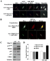
ARF induces nucleolar localization and down regulation of Mip130/LIN-9. The subcellular localization of Mip130/LIN-9 was assessed by confocal microscopy using NIH3T3 cells co-transfected with GFP- or myc-tagged versions of Mip130/LIN-9 (A and B, respectively) and RFP-ARF or RFP alone. The B23/Nucleophosmin protein was used a nucleolar marker. Nuclei were stained with Hoechst. C) The protein expression of Mip130/LIN-9 and components of the ARF-p53 pathway (ARF, p53, MDM2 and p21) was determined by Western blotting with specific antibodies. The expression of CDK1 was used as marker for the expression of G2/M genes known to be downregulated by ARF and the decrease in Mip130/LIN-9. D) ARF does not affect the transcription of Mip130/LIN-9. NIH3T3 cells were transfected with GFP or GFP-ARF and the expression of the Mip130/LIN-9 mRNA was assessed by RT-PCR as described in Materials and Methods. The levels of MDM-2 mRNA, whose transcription is enhanced by ARF via the activation of p53, was used as a positive control.
Figure 3
Figure 3
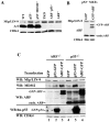
The requirement of p53 in the ARF-induced decrease in Mip130/LIN-9 expression. A) Expression of Mip130/LIN-9 in MEFs deficient for the different genes involved in the ARF-MDM2-p53 pathway. MEFs derived from mice lacking p53, p53 and MDM2, Arf, Arf and p53, all 3 genes (TKO), and wild type were assessed for the expression of Mip130/LIN-9 by Western blot as described in Materials and Methods. The expression of ARF and CDK4 (control loading) was also determined using specific antibodies (see Materials and Methods). B) The expression of ARF does not affect Mip130/LIN-9 in p53-/- MEFs. MEFs null for p53 were transfected with GFP-ARF or control GFP and immunoblotted with Mip130/LIN-9, ARF, and CDK4 antibodies. C) Arf-/- or p53-/- MEFs were transfected with plasmids encoding GFP, GFP-ARF, or GFP-p53. Immunoblotting was performed with antibodies against ARF, human p53 (DO-1), CDK4 or Mip130/LIN-9. The migration of endogenous ARF (endo. ARF) and GFP-ARF is indicated.
Figure 4
Figure 4
ARF induces the degradation of the Mip130/LIN-9 protein through a proteasome-independent pathway. A) The effect of ARF on the half-life of the Mip130/LIN-9 protein was assessed by Western blot with the anti- Mip130/LIN-9 mAb#6 at different time points after cycloheximide addition to the cultures. The level of the Mip130/LIN-9 protein detected was normalized by the level of CDK4, and plotted to show the accelerated decay of this protein in cells expressing ARF. Intensity of the bands was determined using the image processing software program ImageJ. Results are expressed as the mean ± standard deviation obtained from 4 different experiments. B) The effect of ARF and p53 on Mip130/LIN-9 expression in the presence of the proteasome inhibitor MG132 (MG) was assessed in NIH3T3 cells 24 hours after transfection by Western blotting. The expression of MDM2, p21, and p53 were used as control for the effect of MG-132, which is known to increase their expression through the inhibition of the proteasome.
Figure 5
Figure 5
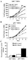
The knockdown of Mip130/LIN-9 inhibits cell proliferation and induces a senescence-like state in p53-/- MEFs. A and B) Arf-/- or p53-/- MEFs were infected with empty retrovirus or encoding the Mip130/LIN-9 shRNA. Cells were seeded at 10,000/well and counted every day in duplicate for 6 days as previously described for cells expressing a Mip130/LIN-9 antisense (Pilkinton et al., 2007b). C) The expression of SA-ß-galactosidase (ß-Gal) was performed as described in Materials and Methods and expressed as the mean ± standard deviation of two separate experiments using duplicate samples.
Figure 6
Figure 6
Mip130/LIN-9 knockdown induces the expression of p16 and ARF. p53-/- and Arf-/- MEF stable clones expressing an shRNA (A and C, respectively) or an antisense (B) against Mip130/LIN-9 were studied for the expression of the indicated senescence markers by Western blotting. D) The expression of the pockets proteins and phospho-RB795 were determined in the p53-/- MEFs with a knockdown of Mip130/LIN-9. E) The levels of mRNA encoding p16 and ARF were determined by RT-PCR in p53-/- MEFs expressing antisense against Mip130/LIN-9 or pBabe-puro vector.
Figure 7
Figure 7
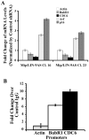
The knockdown of Mip130/LIN-9 inhibited the expression of CDC6 and BubR1 while enhancing the transcription of the Ink4a/Arf locus products, p16 and ARF. A) The mRNA expression for the indicated genes was determined using quantitative real-time PCR as described in Materials and Methods in two Mip130/LIN-9 antisense clones and compared with control clones expressing empty vector. Message levels were normalized by the expression of GAPDH and the effect of the Mip130/LIN-9 antisense was expressed as fold change over control empty vector. B) The direct binding of Mip130/LIN-9 to the cdc6 and BubR1 gene promoters was tested by ChIP assays with specific anti- Mip130/LIN-9 antibodies and primers for the specific amplification of the BubR1 and cdc6 promoters.
Figure 8
Figure 8
The induction of ARF leads to the degradation of Mip130/LIN-9 by a process that is p53-dependent. The degradation of Mip130/LIN-9 protein has two effects. First, B-Myb becomes degraded, which results in a lack of G2-phase gene induction and this contributes to cell cycle arrest. Second, the decrease in Mip130/LIN-9 leads to a decrease in the transcription of negative regulators of the Ink4a/Arf locus, BUBR1 and CDC6, and a subsequent increase in ARF and p16 expression.

Similar articles

Cited by

References

    1. Baker DJ, Jeganathan KB, Malureanu L, Perez-Terzic C. Terzic A and van Deursen JM. J Cell Biol. 2006;172:529–40. - PMC - PubMed
    1. Baker DJ, Jin F, van Deursen JM. Cell Cycle. 2008a;7:2795–802. - PMC - PubMed
    1. Baker DJ, Perez-Terzic C, Jin F, Pitel K, Niederlander NJ, Jeganathan K, Yamada S, Reyes S, Rowe L, Hiddinga HJ, Eberhardt NL, Terzic A, van Deursen JM. Nat Cell Biol. 2008b;10:825–36. - PMC - PubMed
    1. Bates S, Phillips AC, Clark PA, Stott F, Peters G, Ludwig RL, Vousden KH. Nature. 1998;395:124–5. - PubMed
    1. Beijersbergen RL, Carlee L, Kerkhoven RM, Bernards R. Genes Dev. 1995;9:1340–53. - PubMed

Publication types

Substances