

Clear cell sarcoma: a case mimicking primary cutaneous malignant melanoma
- PMID: 20101313
- PMCID: PMC2807157
- DOI: 10.4103/0019-5154.53193
Clear cell sarcoma: a case mimicking primary cutaneous malignant melanoma
Abstract
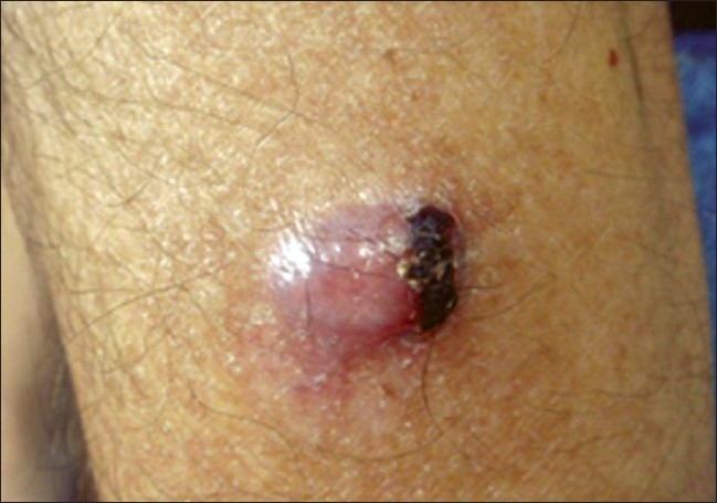
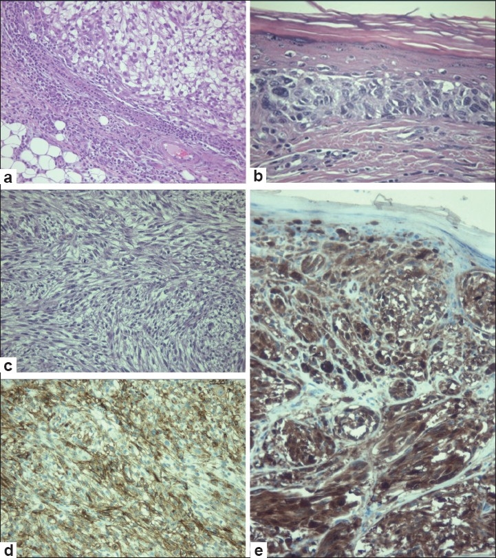
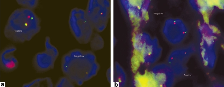
Clear cell sarcoma (CCS) is a recently described variant of sarcoma characterized by prominent clear cells showing features similar to clear cell melanoma. This neoplasm was first described by Dr. Franz M. Erzinger. Primary CCS usually arises in deeper soft tissues, in association with fascia, tendons, or aponeuroses. Characteristic translocation t(12;22) (q13;q12) has been considered pathognomonic for CCS. Prognosis is related to tumor size. An early recognition and initial radical surgery is the key to a favourable outcome. We present a patient with an unusual neoplasm that resembled malignant melanoma.
Keywords: Clear cell sarcoma; genetic studies; malignant melanoma.
Conflict of interest statement
Figures
Similar articles
-
Lessons to be learned: a case study approach. Malignant melanoma of soft tissue.J R Soc Promot Health. 2005 Jan;125(1):42-6. doi: 10.1177/146642400512500114. J R Soc Promot Health. 2005. PMID: 15712852
-
Clear cell sarcoma of tendons and aponeuroses with t(12;22) (q13;q12) diagnosed initially as malignant melanoma.Cancer Genet Cytogenet. 1996 Oct 1;91(1):37-9. doi: 10.1016/s0165-4608(96)00113-6. Cancer Genet Cytogenet. 1996. PMID: 8908164
-
Gastrointestinal melanoma or clear cell sarcoma? Molecular evaluation of 7 cases previously diagnosed as malignant melanoma.Am J Surg Pathol. 2008 Jun;32(6):858-66. doi: 10.1097/PAS.0b013e31815b8288. Am J Surg Pathol. 2008. PMID: 18408594
-
Clear Cell Sarcoma (CCS) of the Soft Tissue: An Update Narrative Review with Emphasis on the Utility of PRAME in Differential Diagnosis.J Clin Med. 2025 Feb 13;14(4):1233. doi: 10.3390/jcm14041233. J Clin Med. 2025. PMID: 40004764 Free PMC article. Review.
-
Translocation t(12;22)(q13;q12.2-12.3) in a clear cell sarcoma of tendons and aponeuroses.Genes Chromosomes Cancer. 1993 Apr;6(4):249-52. doi: 10.1002/gcc.2870060412. Genes Chromosomes Cancer. 1993. PMID: 7685631 Review.
Cited by
-
Intestinal Clear Cell Sarcoma-A Case Presentation of an Extremely Rare Tumor and Literature Review.Medicina (Kaunas). 2024 May 22;60(6):847. doi: 10.3390/medicina60060847. Medicina (Kaunas). 2024. PMID: 38929464 Free PMC article. Review.
-
Clear Cell Sarcoma Incidence and Survival: A Surveillance, Epidemiology, and End Results (SEER) Database Analysis.Cureus. 2025 Jun 9;17(6):e85606. doi: 10.7759/cureus.85606. eCollection 2025 Jun. Cureus. 2025. PMID: 40636612 Free PMC article.
-
Soft Tissue Sarcoma Mimicking Melanoma: A Systematic Review.Cancers (Basel). 2023 Jul 12;15(14):3584. doi: 10.3390/cancers15143584. Cancers (Basel). 2023. PMID: 37509250 Free PMC article. Review.
-
A Comparison of Clear Cell Sarcoma to Jaw and Salivary Tumors Bearing EWS Fusions.Head Neck Pathol. 2024 Mar 25;18(1):25. doi: 10.1007/s12105-024-01625-6. Head Neck Pathol. 2024. PMID: 38526767 Free PMC article. Review.
-
Cutaneous clear cell sarcoma: a rare aggressive tumor with potential diagnostic challenge.J Lab Physicians. 2012 Jan;4(1):53-5. doi: 10.4103/0974-2727.98677. J Lab Physicians. 2012. PMID: 22923926 Free PMC article.
References
-
- Dewan M, Malatani TS, Ansari MA. Lessons to be learned: A case study approach Malignant melanoma of soft tissue. J R Soc Health. 2005;125:42–6. - PubMed
-
- Crowson A, Magro C, Mihm M. Unusual histologic and clinical variants of melanoma: Implications for therapy. Curr Oncol Rep. 2007;9:403–10. - PubMed
-
- Lucas DR, Nascimento AG, Sim FH. Cell sarcoma of soft tissues: Mayo Clinic experience with 35 cases. Am J Surg Pathol. 1992;16:1197–204. - PubMed
-
- Enzinger FM. Clear cell sarcoma of tendons and aponeuroses: An analysis of 21 cases. Cancer. 1965;18:1163–76. - PubMed
-
- Kawai A, Hosono A, Nakayama R, Matsumine A, Matsumoto S, Ueda T, et al. Clear cell sarcoma of tendons and aponeuroses A study of patients. Cancer. 2007;109:109–16. - PubMed
LinkOut - more resources
Full Text Sources