

Vitiligo and the melanocyte reservoir
- PMID: 20101329
- PMCID: PMC2807704
- DOI: 10.4103/0019-5154.57604
Vitiligo and the melanocyte reservoir
Abstract
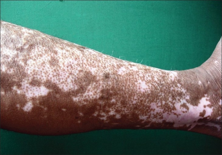
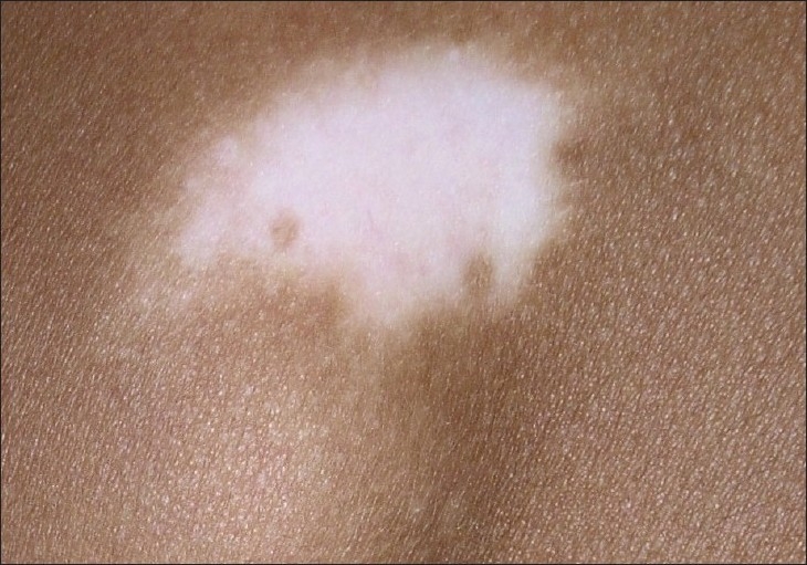
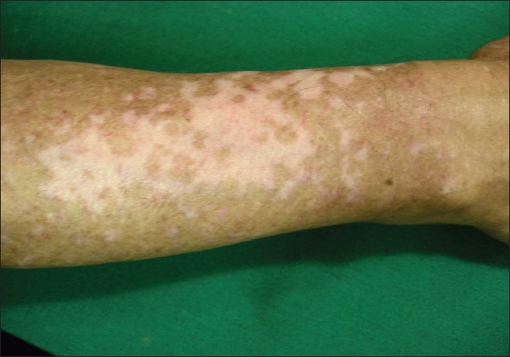
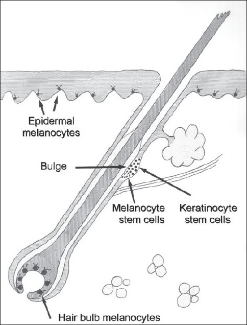
Repigmentation of vitiligo depends on available melanocytes from three possible sources: from the hair follicle unit which is the main provider of pigment cells, from the border of vitiligo lesions, and from unaffected melanocytes within depigmented areas; pigment cells at these locations originate a perifollicular, border spreading and a diffuse repigmentation pattern. In order for repigmentation to take place under stimulation with diverse therapies, melanocytes should be present in appropriate numbers. Melanocyte tissue stem cells located in the niche at the bulge region of the hair follicle are the most important sources for providing immature pigment cells that undergo terminal differentiation and originate repigmentation, but cytokines, UVR and other molecules acting in melanogenesis with adequate regulation mechanisms contribute to successful recovery in vitiligo. The presence of keratinocyte stem cells in the interfollicular epidermis raises the question on the possibility of melanocyte stem cells in a similar location and the development of future strategies for therapeutic purposes.
Keywords: Hair follicle; melanocyte; reservoir; stem cells; vitiligo.
Conflict of interest statement
Figures
Similar articles
-
Repigmentation through Melanocyte Regeneration in Vitiligo.Dermatol Clin. 2017 Apr;35(2):205-218. doi: 10.1016/j.det.2016.11.015. Dermatol Clin. 2017. PMID: 28317529 Review.
-
Skin pigmentation and its control: From ultraviolet radiation to stem cells.Exp Dermatol. 2021 Apr;30(4):560-571. doi: 10.1111/exd.14260. Epub 2020 Dec 24. Exp Dermatol. 2021. PMID: 33320376 Free PMC article. Review.
-
Mechanisms of repigmentation induced by photobiomodulation therapy in vitiligo.Exp Dermatol. 2019 Feb;28 Suppl 1:10-14. doi: 10.1111/exd.13823. Exp Dermatol. 2019. PMID: 30698884 Review.
-
Repigmentation of Human Vitiligo Skin by NBUVB Is Controlled by Transcription of GLI1 and Activation of the β-Catenin Pathway in the Hair Follicle Bulge Stem Cells.J Invest Dermatol. 2018 Mar;138(3):657-668. doi: 10.1016/j.jid.2017.09.040. Epub 2017 Oct 17. J Invest Dermatol. 2018. PMID: 29054607 Free PMC article.
-
Melanocyte Precursors in the Hair Follicle Bulge of Repigmented Vitiligo Skin Are Controlled by RHO-GTPase, KCTD10, and CTNNB1 Signaling.J Invest Dermatol. 2021 Mar;141(3):638-647.e13. doi: 10.1016/j.jid.2020.07.016. Epub 2020 Aug 13. J Invest Dermatol. 2021. PMID: 32800877
Cited by
-
The Role of Memory CD8+ T Cells in Vitiligo.J Immunol. 2019 Jul 1;203(1):11-19. doi: 10.4049/jimmunol.1900027. J Immunol. 2019. PMID: 31209143 Free PMC article. Review.
-
From neural crest cells to melanocytes: cellular plasticity during development and beyond.Cell Mol Life Sci. 2019 May;76(10):1919-1934. doi: 10.1007/s00018-019-03049-w. Epub 2019 Mar 4. Cell Mol Life Sci. 2019. PMID: 30830237 Free PMC article. Review.
-
Dermatological Conditions in SKIN OF COLOR-: Debunking Vitiligo Myths.J Clin Aesthet Dermatol. 2022 Sep;15(9 Suppl 1):S10-S11. J Clin Aesthet Dermatol. 2022. PMID: 36118789 Free PMC article. No abstract available.
-
[Treatment of vitiligo].Hautarzt. 2016 Mar;67(3):249-64. doi: 10.1007/s00105-016-3768-4. Hautarzt. 2016. PMID: 26909811 German.
-
Deciphering skin re-pigmentation patterns in vitiligo: an update on the cellular and molecular events involved.Chin Med J (Engl). 2020 May 20;133(10):1231-1238. doi: 10.1097/CM9.0000000000000794. Chin Med J (Engl). 2020. PMID: 32433056 Free PMC article. Review.
References
-
- Nordlund JJ, Ortonne JP. Vitiligo vulgaris. In: Nordlund JJ, Boissy RE, Hearing VJ, King RA, Ortonne JP, editors. The pigmentary system: Physiology and pathophysiology. New York: Oxford University Press; 1998. pp. 513–51.
-
- Falabella R. Surgical treatment of vitiligo: Why, when and how. J Eur Acad Dermatol Venereol. 2003;17:518–20. - PubMed
-
- Falabella R, Barona MI. Update on skin repigmentation therapies in vitiligo. Pigment Cell Melanoma Res. 2009;22:42–65. - PubMed
-
- Parsad D, Pandhi R, Dogra S, Kumar B. Clinical study of repigmentation patterns with different treatment modalities and their correlation with speed and stability of repigmentation in 352 vitiliginous patches. J Am Acad Dermatol. 2004;50:63–7. - PubMed
LinkOut - more resources
Full Text Sources