

Quantitation of mast cells and collagen fibers in skin tags
- PMID: 20101330
- PMCID: PMC2807705
- DOI: 10.4103/0019-5154.57605
Quantitation of mast cells and collagen fibers in skin tags
Abstract
Background: Skin tags are common benign skin tumors usually occurring on the neck and major flexors of elder people.
Aims: The aim of this study is to perform quantitation of mast cells and collagen fibers in skin tags and normal skin in diabetics and nondiabetics, to find a possible correlation between mast cells and collagen fibers in the pathogenesis of skin tags.
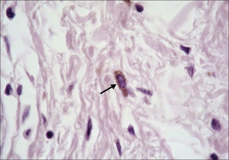
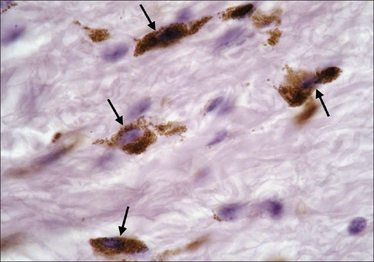
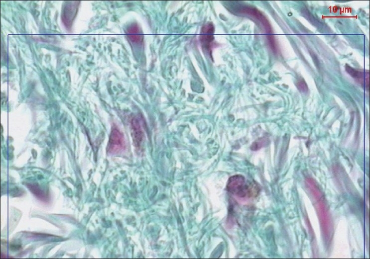
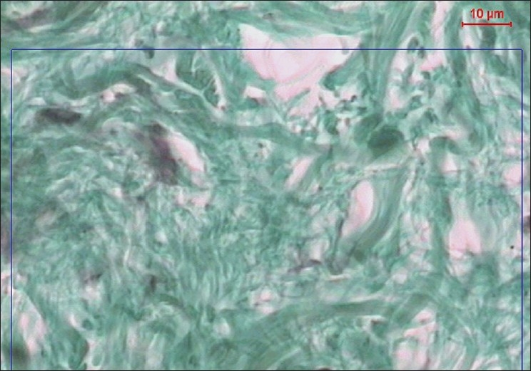
Methods: Thirty participants with skin tags were divided into two groups (15 diabetic and 15 nondiabetic). Three biopsies were obtained from one anatomical site: A large skin tag, a small skin tag, and adjacent normal skin. Mast cells stained with Bismarck brown were counted manually in ten different fields of each section with magnification x1000 and the average count was correlated with the percentage of mean collagen area in five fields done by the image analyzer.
Results: A statistically significant correlation between mast cell count and percentage of collagen mean area was detected in both studied groups (except in large skin tags of the nondiabetic group).
Conclusion: The positive correlation between mast cell count and percentage of collagen mean area suggests the critical role of mast cells in the etiogenesis of skin tags through its interaction with fibroblasts.
Keywords: Bismarck brown; Image analyzer; mast cells; mean collagen area %; skin tags.
Conflict of interest statement
Figures
Similar articles
-
Skin tag as a cutaneous marker for impaired carbohydrate metabolism: a case-control study.Int J Dermatol. 2007 Nov;46(11):1155-9. doi: 10.1111/j.1365-4632.2007.03287.x. Int J Dermatol. 2007. PMID: 17988334
-
Role of mast cells in skin tag development: an immunohistochemical study.Anal Quant Cytopathol Histpathol. 2014 Aug;36(4):222-30. Anal Quant Cytopathol Histpathol. 2014. PMID: 25291860
-
A study of androgen and estrogen receptors alpha, beta in skin tags.Indian J Dermatol. 2010;55(1):20-4. doi: 10.4103/0019-5154.60345. Indian J Dermatol. 2010. PMID: 20418971 Free PMC article.
-
Age related changes in the dermal mast cells and the associated changes in the dermal collagen and cells: A histological and electron microscopy study.Acta Histochem. 2019 Jul;121(5):619-627. doi: 10.1016/j.acthis.2019.05.004. Epub 2019 May 22. Acta Histochem. 2019. PMID: 31126613
-
The possible role of trauma in skin tags through the release of mast cell mediators.Indian J Dermatol. 2011 Nov;56(6):641-6. doi: 10.4103/0019-5154.91819. Indian J Dermatol. 2011. PMID: 22345761 Free PMC article.
Cited by
-
A clinical evaluation of skin tags in relation to obesity, type 2 diabetis mellitus, age, and sex.Indian J Dermatol. 2011 Jul;56(4):393-7. doi: 10.4103/0019-5154.84765. Indian J Dermatol. 2011. PMID: 21965846 Free PMC article.
-
Skin tags: a link between lesional mast cell count/tryptase expression and obesity and dyslipidemia.Indian J Dermatol. 2013 May;58(3):240. doi: 10.4103/0019-5154.110843. Indian J Dermatol. 2013. PMID: 23723485 Free PMC article.
-
Subcutaneous Injection and Brush Application of Ovalbumin-Aluminum Salt Solution Induces Dermatitis-like Changes in Mice.J Clin Med. 2025 Mar 3;14(5):1701. doi: 10.3390/jcm14051701. J Clin Med. 2025. PMID: 40095628 Free PMC article.
-
Interplay of collagen and mast cells in periapical granulomas and periapical cysts: a comparative polarizing microscopic and immunohistochemical study.Restor Dent Endod. 2022 Feb 14;47(1):e12. doi: 10.5395/rde.2022.47.e12. eCollection 2022 Feb. Restor Dent Endod. 2022. PMID: 35284324 Free PMC article.
References
-
- Chiritescu E, Maloney ME. Acrochordons as a presenting sign of nevoid basal cell carcinoma syndrome. J Am Acad Dermatol. 2001;44:789–94. - PubMed
-
- Almeida SC, Perez PM, Sánchez AT. Giant skin tags: Report of two cases. Dermatol Online J. 2007;13:30. - PubMed
-
- Kahana M, Grossman E, Feinstein A, Cohen M, Ronnen M, Millet MS. Skin tags: A cutaneous marker for diabetes mellitus. Acta Dermatol Venereol. 1987;67:175–7. - PubMed
-
- Garcia-Hidalgo L, Orozco-Tropete R, Villa AR, Dalman JJ. Dermatoses in 156 obese adults. Obes Res. 1999;7:299–302. - PubMed
-
- Ben-Shlomo A, Melmed S. Skin manifestations in acromegaly. Clin Dermatol. 2006;24:256–9. - PubMed
LinkOut - more resources
Full Text Sources