

White-matter abnormalities in brain during early abstinence from methamphetamine abuse
- PMID: 20101394
- PMCID: PMC2819660
- DOI: 10.1007/s00213-009-1761-7
White-matter abnormalities in brain during early abstinence from methamphetamine abuse
Abstract
Background: Previous studies revealed microstructural abnormalities in prefrontal white matter and corpus callosum of long-term abstinent chronic methamphetamine abusers. In view of the importance of the early abstinence period in treatment retention, we compared 23 methamphetamine-dependent subjects abstinent from methamphetamine for 7-13 days with 18 healthy comparison subjects. As certain metabolic changes in the brain first manifest after early abstinence from methamphetamine, it is also possible that microstructural white-matter abnormalities are not yet present during early abstinence.
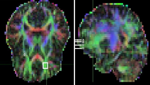
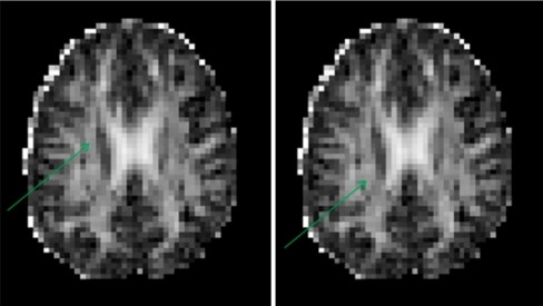
Methods: Using diffusion tensor imaging at 1.5 T, fractional anisotropy (FA) was measured in prefrontal white matter at four inferior-superior levels parallel to the anterior commissure-posterior commissure (AC-PC) plane. We also sampled FA in the corpus callosum at the midline and at eight bilateral, fiber-tract sites in other regions implicated in effects of methamphetamine.
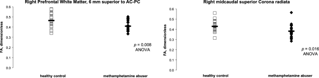
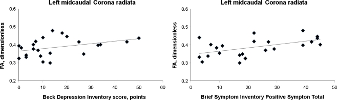
Results: The methamphetamine group exhibited lower FA in right prefrontal white matter above the AC-PC plane (11.9% lower; p = 0.007), in midline genu corpus callosum (3.9%; p = 0.019), in left and right midcaudal superior corona radiata (11.0% in both hemispheres, p's = 0.020 and 0.016, respectively), and in right perforant fibers (7.3%; p = 0.025). FA in left midcaudal superior corona radiata was correlated with depressive and generalized psychiatric symptoms within the methamphetamine group.
Conclusions: The findings support the idea that methamphetamine abuse produces microstructural abnormalities in white matter underlying and interconnecting prefrontal cortices and hippocampal formation. These effects are already present during the first weeks of abstinence from methamphetamine and are linked to psychiatric symptoms assessed during this period.
Figures

Similar articles
-
Reduced corpus callosum white matter microstructural integrity revealed by diffusion tensor eigenvalues in abstinent methamphetamine addicts.Neurotoxicology. 2009 Mar;30(2):209-13. doi: 10.1016/j.neuro.2008.12.002. Epub 2008 Dec 24. Neurotoxicology. 2009. PMID: 19135475
-
Decreased frontal white-matter integrity in abstinent methamphetamine abusers.Int J Neuropsychopharmacol. 2007 Dec;10(6):765-75. doi: 10.1017/S1461145706007395. Epub 2006 Dec 6. Int J Neuropsychopharmacol. 2007. PMID: 17147837
-
Cognitive control and white matter callosal microstructure in methamphetamine-dependent subjects: a diffusion tensor imaging study.Biol Psychiatry. 2009 Jan 15;65(2):122-8. doi: 10.1016/j.biopsych.2008.08.004. Epub 2008 Sep 23. Biol Psychiatry. 2009. PMID: 18814867 Free PMC article.
-
[The value of MR diffusion tensor imaging in assessing white matter changes in short-term methamphetamine withdrawal].Zhonghua Yi Xue Za Zhi. 2022 Sep 20;102(35):2779-2785. doi: 10.3760/cma.j.cn112137-20220113-00091. Zhonghua Yi Xue Za Zhi. 2022. PMID: 36124350 Chinese.
-
Abuse of amphetamines and structural abnormalities in the brain.Ann N Y Acad Sci. 2008 Oct;1141:195-220. doi: 10.1196/annals.1441.031. Ann N Y Acad Sci. 2008. PMID: 18991959 Free PMC article. Review.
Cited by
-
Cigarette smoking and white matter microstructure.Psychopharmacology (Berl). 2012 May;221(2):285-95. doi: 10.1007/s00213-011-2621-9. Epub 2012 Jan 4. Psychopharmacology (Berl). 2012. PMID: 22215225 Free PMC article.
-
Striatum and insula dysfunction during reinforcement learning differentiates abstinent and relapsed methamphetamine-dependent individuals.Addiction. 2014 Mar;109(3):460-71. doi: 10.1111/add.12403. Epub 2013 Dec 15. Addiction. 2014. PMID: 24329936 Free PMC article.
-
White Matter Abnormalities Based on TBSS and Its Correlation With Impulsivity Behavior of Methamphetamine Addicts.Front Psychiatry. 2020 May 21;11:452. doi: 10.3389/fpsyt.2020.00452. eCollection 2020. Front Psychiatry. 2020. PMID: 32528325 Free PMC article.
-
Chronic methamphetamine abuse and corticostriatal deficits revealed by neuroimaging.Brain Res. 2015 Dec 2;1628(Pt A):174-85. doi: 10.1016/j.brainres.2014.10.044. Epub 2014 Nov 4. Brain Res. 2015. PMID: 25451127 Free PMC article. Review.
-
Aberrant topology of white matter networks in patients with methamphetamine dependence and its application in support vector machine-based classification.Sci Rep. 2023 Apr 28;13(1):6958. doi: 10.1038/s41598-023-33199-8. Sci Rep. 2023. PMID: 37117256 Free PMC article.
References
Publication types
MeSH terms
Substances
Grants and funding
LinkOut - more resources
Full Text Sources
Medical
Molecular Biology Databases
Research Materials
