Review
. 2010 Jun;67(11):1779-98.
doi: 10.1007/s00018-010-0259-0. Epub 2010 Jan 27.

Protein folding in membranes

Affiliations
Review

Protein folding in membranes

Sebastian Fiedler et al. Cell Mol Life Sci. 2010 Jun.

Abstract

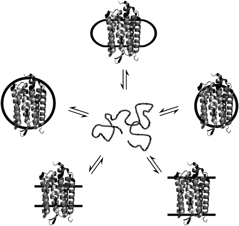
Separation of cells and organelles by bilayer membranes is a fundamental principle of life. Cellular membranes contain a baffling variety of proteins, which fulfil vital functions as receptors and signal transducers, channels and transporters, motors and anchors. The vast majority of membrane-bound proteins contain bundles of alpha-helical transmembrane domains. Understanding how these proteins adopt their native, biologically active structures in the complex milieu of a membrane is therefore a major challenge in today's life sciences. Here, we review recent progress in the folding, unfolding and refolding of alpha-helical membrane proteins and compare the molecular interactions that stabilise proteins in lipid bilayers. We also provide a critical discussion of a detergent denaturation assay that is increasingly used to determine membrane-protein stability but is not devoid of conceptual difficulties.


Figures

Fig. 1
Fig. 1
Seven-helix phototaxis receptor sensory rhodopsin II in a phospholipid bilayer. A high-resolution protein structure (PDB 1jgj) obtained by X-ray crystallography [182] was superimposed onto a bilayer manually assembled from 3,648 1-palmitoyl-2-oleoyl-sn-glycero-phosphocholine (POPC, Molfile from Avanti Polar Lipids, Alabaster, AL) monomers. Each of the two headgroup regions is about 15 Å thick, whereas the hydrocarbon core is about 30 Å in thickness
Fig. 2
Fig. 2
Schematic representation of water-soluble protein folding, exemplified using human carbonyl reductase 1 (276 residues). In an idealised scenario, a completely unfolded state devoid of specific intramolecular interactions and stable structure (left) is in equilibrium with a folded, biologically active state (right; PDB 1wma). Note that the unfolded states of several proteins have been shown to retain considerable amounts of secondary structure and long-range contacts [181] and that folding and unfolding reactions may involve kinetic and equilibrium intermediates [183]. In both panels, hydrophilic amino acid residues (Arg, Asn, Asp, Gln, Glu, His, Lys) are shown in black, whereas others are coloured grey
Fig. 3
Fig. 3
Specific interactions in water-soluble protein folding. Colour code for residues shown in atomic detail is red for oxygen, blue for nitrogen and white for hydrogen. a Interhelical hydrogen bonds. The homodimer of the GCN4-leucine zipper from Saccharomyces cerevisiae (PDB 2zta) contains an interhelical Asn16–Asn16 (orange) hydrogen bond (dashed line). b van der Waals contacts. Large side chains of helices 7 (orange) and 8 (yellow) interdigitate to form extensive van der Waals contacts in E. coli J-type co-chaperone HSC20 (PDB 1fpo). This example illustrates also the burial of hydrophobic side chains from water, which is a manifestation of the hydrophobic effect. c Aromatic-aromatic interactions. Two Phe10–Phe29 pairs are involved in π–π interactions in the homodimeric de novo-designed protein α2D (PDB 1qp6). d Salt bridges. A salt bridge encompassing His31 (green) and Asp70 (yellow) stabilises T4 lysozyme (PDB 2lzm)
Fig. 4
Fig. 4
Differences in the hydrophobicity of surface areas between globular water-soluble and α-helical membrane proteins of similar size. Colour code is blue for hydrophilic surfaces, orange for hydrophobic ones and dark salmon for residues that are in between. a Human carbonyl reductase 1 (PDB 1wma, 276 residues) presents mainly hydrophilic residues to its aqueous environment and shields hydrophobic ones in its core. b Bacteriorhodopsin from Halobacterium salinarum (PDB 1c3w, 231 residues) exposes mostly hydrophobic residues to its lipid bilayer environment, whereas hydrophilic residues are found in the rather small regions that are in contact with lipid headgroups and water. Note that the interiors of membrane proteins are as hydrophobic as those of water-soluble proteins (not shown, see [20, 22])
Fig. 5
Fig. 5
Schematic representation of the two-stage model of membrane-protein folding, exemplified using bacteriorhodopsin from Halobacterium salinarum (231 residues). In stage 1, an unfolded polypeptide chain (top) is inserted into a lipid bilayer to form a loose, biologically inactive bundle rich in α-helical secondary structure (bottom left). In stage 2, interhelical interactions give rise to a compactly folded, biologically active state (bottom right, PDB 1c3w). Note that a water-soluble unfolded state (top) does not exist for most α-helical membrane proteins (see “Determining the stability of α-helical membrane proteins”). In these cases, stage 1 corresponds to the translocon-mediated membrane insertion of nascent polypeptide chains. Also, the depiction of the bilayer-bound denatured state (bottom left) is schematic, as no high-resolution structure is available. In all panels, hydrophilic amino acid residues (Arg, Asn, Asp, Gln, Glu, His, Lys) are shown in black, whereas others are coloured grey
Fig. 6
Fig. 6
Specific interactions in membrane-protein folding. Colour code for residues shown in atomic detail is red for oxygen, blue for nitrogen and white for hydrogen. a Interhelical hydrogen bonds. In bacteriorhodopsin from Halobacterium salinarum (PDB 1c3w), Tyr185 (orange) of helix 6 and Asp212 (yellow) of helix 7 form an interhelical hydrogen bond (dashed line). b van der Waals contacts. Small residues (orange and yellow) increase the homodimer interface and allow for extensive van der Waals contacts in human glycophorin A (PDB 1afo). Hydrogens are shown for Gly residues only. c Aromatic–aromatic interactions. In subunit I of aberrant ba 3-cytochrome c oxidase from Thermus thermophilus (PDB 1ehk), Trp110 of helix 4 interacts with Tyr23 and Leu27 of helix 1, although it is partly exposed to the lipid bilayer. d Salt bridges. Lys358 and Asp237 are crucial for membrane insertion of lac permease from E. coli (PDB 2v8n) and have been suggested to form a salt bridge within the protein’s transmembrane region [95, 96]
Fig. 7
Fig. 7
Dependence of protein stability on cavity surface area in water-soluble and membrane proteins. Experimental data for T4 lysozyme (open squares, taken from [80]) and bacteriorhodopsin (filled circles, [79]) as well as linear regressions (dash-dotted and solid lines, respectively). The stabilities of both proteins decreased in a roughly linear fashion with an increase in cavity surface area caused by the large-to-small amino acid substitutions indicated in the figure. Similar slopes observed for the two proteins suggest that van der Waals forces contribute equally to the stabilities of water-soluble and membrane proteins. The y-axis intercepts are different because of the greater contribution of the hydrophobic effect in thermal unfolding of T4 lysozyme as compared with SDS denaturation of bacteriorhodopsin. Figure adapted with permission from [79]. Copyright 2009 American Chemical Society
Fig. 8
Fig. 8
Schematic phospholipid shapes (left) and lateral pressure profiles (right) in bilayers composed of those lipids. Lateral pressure, p, is plotted versus bilayer depth, z. a In “cylindrical” lipids, like POPC, the headgroup and the acyl chains have similar area requirements. b Addition of lipids with small headgroups, such as POPE, redistributes positive lateral pressure from the headgroup regions to the hydrocarbon core. c Vice versa, addition of lipids with only one acyl chain (lysolipids), such as lysoPC, lowers the positive lateral pressure in the hydrocarbon core but augments it in the headgroup regions. The negative lateral pressure component is due to interfacial tension between the lipids’ acyl chains and their hydrated headgroups and thus remains virtually constant. At equilibrium, the pressure integral across the bilayer is always zero, i.e., the cumulative areas under the p(z) curves are equal for positive and negative pressure components
Fig. 9
Fig. 9
Idealised scenario of an α-helical membrane protein that assumes a monomeric, largely unstructured state in aqueous solution upon reversible unfolding out of different membrane-mimetic systems by a chemical denaturant. Membrane-mimetic systems shown are bicelles (top), micelles of different hydrophobic thicknesses (centre) and lipid bilayers of different hydrophobic thicknesses or lateral pressure profiles (bottom). The protein structure shown is from Halobacterium salinarum bacteriorhodopsin (PDB 1c3w), which was chosen purely for illustrative purposes

Similar articles

Cited by

References

    1. Fersht AR. From the first protein structures to our current knowledge of protein folding: delights and scepticisms. Nat Rev Mol Cell Biol. 2008;9:650–654. doi: 10.1038/nrm2446. - DOI - PubMed
    1. Wallin E, von Heijne G. Genome-wide analysis of integral membrane proteins from eubacterial, archaean, and eukaryotic organisms. Protein Sci. 1998;7:1029–1038. - PMC - PubMed
    1. Overington JP, Al Lazikani B, Hopkins AL. How many drug targets are there? Nat Rev Drug Discov. 2006;5:993–996. doi: 10.1038/nrd2199. - DOI - PubMed
    1. Haber E, Anfinsen CB. Side-chain interactions governing the pairing of half-cystine residues in ribonuclease. J Biol Chem. 1962;237:1839–1844. - PubMed
    1. Anfinsen CB. Principles that govern the folding of protein chains. Science. 1973;181:223–230. doi: 10.1126/science.181.4096.223. - DOI - PubMed

Publication types

LinkOut - more resources