. 2010 Feb;293(2):208-14.
doi: 10.1002/ar.21049.

Proteolytic cleavage of versican during limb joint development

Affiliations

Proteolytic cleavage of versican during limb joint development

Anthony A Capehart. Anat Rec (Hoboken). 2010 Feb.

Abstract

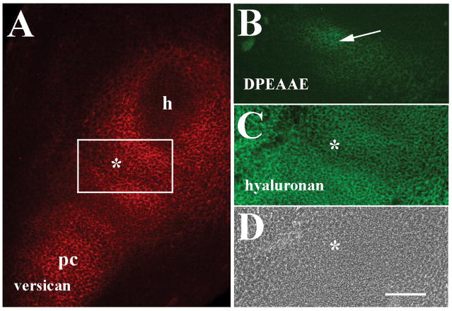
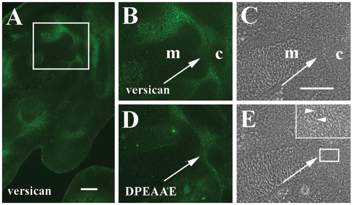
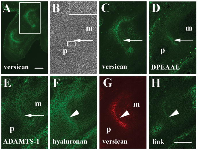
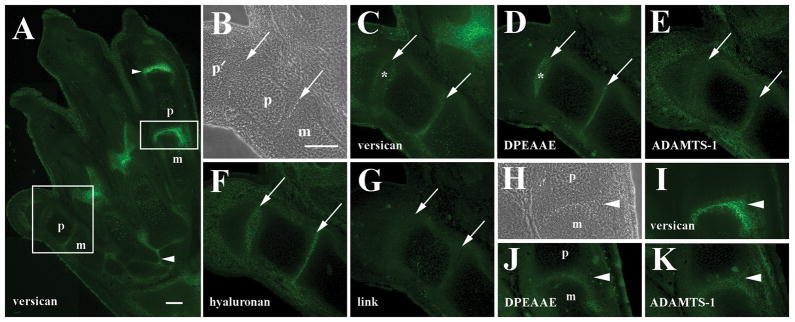
Versican is highly expressed in developing joint interzones during limb morphogenesis. This study was undertaken to examine whether proteolytic cleavage of versican occurs that could potentially impact its function during the process of embryonic synovial joint formation. Using an antibody to the DPEAAE neoepitope generated by ADAMTS proteolysis, versican amino terminal cleavage fragments were detected in joint interzones at 12-16 days post coitum (dpc). ADAMTS-1 localization overlapped that of DPEAAE-reactive versican fragments suggesting it as one possible protease activity involved in processing of versican in the interzone. Results show that increased cleavage of versican in the interzone accompanies cavitation and suggests that proteolytic modification of versican may be important during the process of synovial joint maturation.


Figures

Figure 1
Figure 1
Localization of versican, anti-DPEAAE-reactive versican proteolytic fragments and hyaluronan in the 12 dpc forelimb. A: Versican is localized in the future joint interzone (asterisks in A, C and D) between the humeral primordium (h), and precartilage condensation (pc) of the zeugopod. B: Anti-DPEAAE staining shows low levels of reactivity in the interzone (arrow). C: Double labeling of A shows widespread hyaluronan localization. D: Phase contrast image of A and C. Scale bar = 100 μm for all panels.
Figure 2
Figure 2
Localization of versican and anti-DPEAAE-reactive versican proteolytic fragments in 15 dpc autopod. A: Versican is localized in interzones of carpometacarpal (boxed area) and metacarpophalangeal joints. B: Higher magnification of boxed area in panel A shows versican surrounding interzone cells and around metacarpal edges (m). Note reduction of versican between carpal (c) and metacarpal. C: Phase contrast image of B. D: Anti-DPEAAE cleavage product has overlapping distribution with versican but is also prevalent where versican staining is reduced (arrows B–E). E: Phase contrast image of D. Inset shows higher magnification of boxed area including edge of carpal template and adjacent interzone. Arrowheads indicate separation of some interzone cells. Scale bars = 100 μm in A and C (applies to panels B–E).
Figure 3
Figure 3
Localization of versican, anti-DPEAAE-reactive versican proteolytic fragments, ADAMTS-1, hyaluronan, and link protein in the 15 dpc metacarpophalangeal joint. A: Versican is highly expressed in the interzone of metacarpophalangeal (boxed area) and interphalangeal joints. B: Higher magnification phase contrast image of boxed area in A. Inset shows higher magnification of boxed area including edge of proximal phalanx and adjacent interzone. C: Higher magnification of versican staining in A. D: Anti-DPEAAE reactivity is localized immediately proximal to versican (arrows in B–D). E: ADAMTS-1 staining overlaps that of anti- DPEAAE (arrow). F: Hyaluronan localization in interzone and surrounding tissues. G: Double label of F shows co-distribution of versican with hyaluronan. H: Link protein co-localizes with hyaluronan and versican in the central interzone lamina (arrowheads in F–H). m, metacarpal; p, phalanx. Scale bars = 100μm in A and H (applies to panels B–H).
Figure 4
Figure 4
Localization of versican, anti-DPEAAE-reactive versican proteolytic fragments, ADAMTS-1, and hyaluronan in the 16 dpc metatarsophalangeal joint. A: Versican staining in numerous joints of the hindlimb autopod. Staining is reduced in the metatarsophalangeal joint of digit 1 (larger boxed area) relative to digit 4 (smaller boxed area). Arrowheads, tarsometatarsal and interphalangeal joints. B: Higher magnification phase contrast image of metatarso- and interphalangeal joints (arrows in panels B–G) of digit 1 in A. Cavitation is readily evident. C: Higher magnification of versican staining in A. Versican is reduced in metatarsophalalangeal and interphalangeal interzones. D: Anti-DPEAAE reactivity is localized in the central joint interzone where versican staining is largely absent (asterisks in panels C and D). E: Low level ADAMTS-1 staining in interzones. F: Hyaluronan localization in joint interzones and surrounding tissues. G: Little or no anti-link protein reactivity is observed. H: Higher magnification phase contrast image of metatarsophalangeal joint of digit 4 in A (arrowheads in H–K). I: Higher magnification of versican localization shows strong signal in the interzone. J: Weak anti-DPEAAE reactivity in the joint interzone. K: ADAMTS-1 reactivity in the interzone. m, metatarsal; p and p′, phalanges. Scale bars = 100 μm in A and B (applies to panels B–K).

Similar articles

Cited by

References

    1. Ang LC, Zhang Y, Cao L, Yang BL, Young B, Kiani C, Lee V, Allan K, Yang BB. Versican enhances locomotion of astrocytoma cells and reduces cell adhesion through its G1 domain. J Neuropathol Exp Neurol. 1999;58:597–605. - PubMed
    1. Archer CW, Morrison H, Pitsillides AA. Cellular aspects of the development of diarthroidal joints and articular cartilage. J Anat. 1994;184:447–456. - PMC - PubMed
    1. Craig FM, Bentley G, Archer CW. The spatial and temporal pattern of collagens I and II and keratin sulphate in the developing chick metatarsophalangeal joint. Development. 1987;99:383–391. - PubMed
    1. Craig FM, Bayliss MT, Bentley G, Archer CW. A role for hyaluronan in joint development. J Anat. 1990;171:17–23. - PMC - PubMed
    1. Garciadiego-Cazares D, Rosales C, Katoh M, Cimal-Monroy J. Coordination of chondrocyte differentiation and joint formation by α5β1 integrin in the developing appendicular skeleton. Development. 2004;131:4735–4742. - PubMed

Publication types

LinkOut - more resources