Review
. 2010 Jan-Feb;77(1):32-42.
doi: 10.1002/msj.20157.

Neuropathology of Alzheimer's disease

Affiliations
Review

Neuropathology of Alzheimer's disease

Daniel P Perl. Mt Sinai J Med. 2010 Jan-Feb.

Abstract

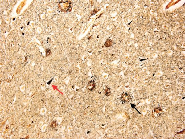
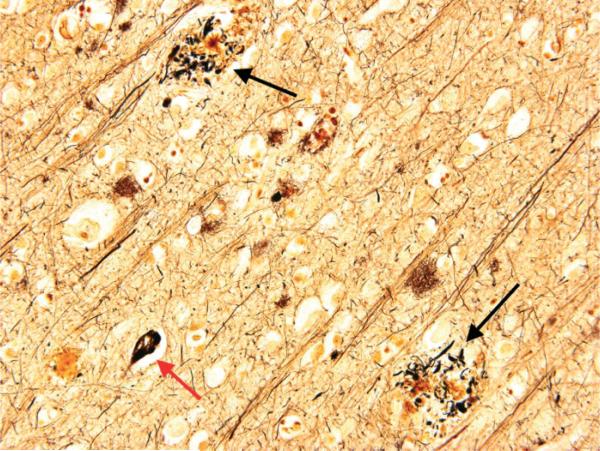
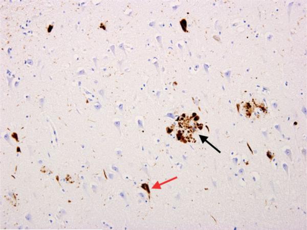
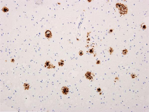
Alois Alzheimer first pointed out that the disease which would later bear his name has a distinct and recognizable neuropathological substrate. Since then, much has been added to our understanding of the pathological lesions associated with the condition. The 2 primary cardinal lesions associated with Alzheimer's disease are the neurofibrillary tangle and the senile plaque. The neurofibrillary tangle consists of abnormal accumulations of abnormally phosphorylated tau within the perikaryal cytoplasm of certain neurons. The senile plaque consists of a central core of beta-amyloid, a 4-kD peptide, surrounded by abnormally configured neuronal processes or neurites. Other neuropathological lesions are encountered in cases of Alzheimer's disease, but the disease is defined and recognized by these 2 cardinal lesions. Other lesions include poorly understood changes such as granulovacuolar degeneration and eosinophilic rodlike bodies (Hirano bodies). The loss of synaptic components is a change that clearly has a significant impact on cognitive function and represents another important morphological alteration. It is important to recognize that distinguishing between Alzheimer's disease, especially in its early stages, and normal aging may be very difficult, particularly if one is examining the brains of patients who died at an advanced old age. It is also noted that instances of pure forms of Alzheimer's disease, in the absence of other coexistent brain disease processes, such as infarctions or Parkinson's disease-related lesions, are relatively uncommon, and this must be taken into account by researchers who employ postmortem brain tissues for research.


Figures

Fig 1
Fig 1
Photomicrograph of the temporal cortex of a patient with Alzheimer's disease (modified Bielschowski stain; original magnification, 40×). Numerous senile (neuritic) plaques (black arrow) and neurofibrillary tangles (red arrow) are shown.
Fig 2
Fig 2
Photomicrograph of the temporal cortex of a patient with Alzheimer's disease (modified Bielschowski stain; original magnification, 100×). Numerous senile (neuritic) plaques (black arrows) and neurofibrillary tangles (red arrow) are shown.
Fig 3
Fig 3
Temporal cortex of a patient with Alzheimer's disease (immunohistochemical stain; original magnification, 100×): the microscopic appearance of an immunohistochemical preparation using an antibody directed against abnormally phosphorylated tau (TG-3; a gift from Dr. P. Davies). This antibody prominently decorates neurofibrillary tangles (red arrow) and swollen dystrophic neurites (neuronal processes) that form the outer rim of the senile (neuritic) plaques (black arrow).
Fig 4
Fig 4
Photomicrograph of the temporal cortex of a patient with Alzheimer's disease (modified Bielschowski stain; original magnification, 400×). Two senile (neuritic) plaques with a neurofibrillary tangle between them are shown.
Fig 5
Fig 5
Temporal cortex of a patient with Alzheimer's disease (immunohistochemical stain; original magnification, 100×): the microscopic appearance of an immunohistochemical preparation using an antibody directed against the components of beta-amyloid (4G8; a gift from Dr. Robakis). This antibody selectively decorates the numerous senile plaques present in this case of advanced Alzheimer's disease and demonstrates the extent of amyloid accumulation that one may encounter in the terminal phases of the disease.

Similar articles

Cited by

References

    1. Terry RD. Interrelations among the lesions of normal and abnormal aging of the brain. Prog Brain Res. 1986;70:41–48. - PubMed
    1. Haroutunian V, Schnaider-Beeri M, Schmeidler J, et al. Role of the neuropathology of Alzheimer disease in dementia in the oldest-old. Arch Neurol. 2008;65:1211–1217. - PMC - PubMed
    1. Alzheimer A. Ueber eine eigenartige erkrankung der hirnrinde. Z Gesamte Neurol Psychiatr. 1907;18:177–179.
    1. Dickson DW, Farlo J, Davies P, et al. Alzheimer's disease. A double-labeling immunohistochemical study of senile plaques. Am J Pathol. 1988;132:86–101. - PMC - PubMed
    1. Yen SH, Dickson DW, Crowe A, et al. Alzheimer's neurofibrillary tangles contain unique epitopes and epitopes in common with the heat-stable microtubule associated proteins tau and MAP2. Am J Pathol. 1987;126:81–91. - PMC - PubMed

Publication types

Substances